|
|
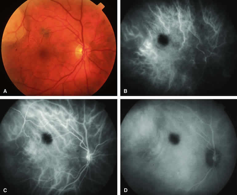
| Fig. 2. Typical melanotic choroidal nevus. A. Small gray choroidal nevus one disc diameter above fovea. The pale lesion at upper left margin of photograph is a choroidal melanoma that was the main focus of the following angiogram. B-D. Indocyanine green (ICG) angiogram of lesion. B. Early-phase frame showing intense hypofluorescence of nevus. C. Intermediate-phase frame showing sustained hypofluorescence of nevus. D. Late-phase frame showing sustained hypofluorescence of nevus. |