|
|
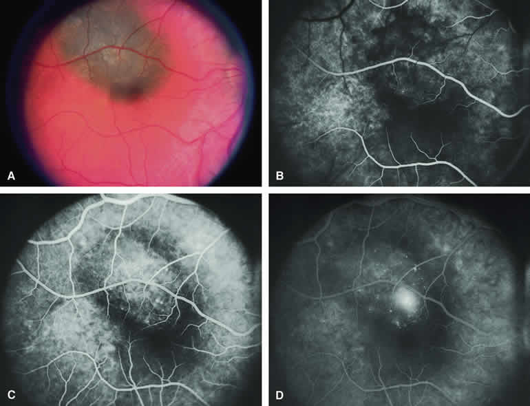
| Fig. 7. Melanotic choroidal nevus versus melanoma with overlying serous subretinal fluid. A. Bland gray-brown choroidal lesion involving upper portion of macula with shallow overlying and surrounding blister of serous subretinal fluid. B-D. Fluorescein angiogram of lesion. B. Arterial phase frame showing patchy choroidal hypofluorescence corresponding to choroidal lesion. C. Full venous phase frame showing faint zone of choroidal hypofluorescence corresponding to marginal zone of lesion, mild hyperfluorescence corresponding to the central portion of the lesion, and discrete pinpoint foci of hyperfluorescence at the retinal pigment epithelial (RPE) level on the lesion. D. Later phase frame showing smudge hyperfluorescent focus of fluorescein leakage into subretinal fluid. |