|
|
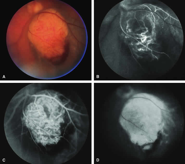
| Fig. 13. Choroidal melanoma with nodular eruption through Bruch's membrane. A. Mushroom-shaped choroidal melanoma inferonasal to optic disc, showing dark basal region and lighter apical eruption through Bruch's membrane. Note prominence of intralesional blood vessels within apical portion of lesion. B-D. Fluorescein angiogram of lesion. B. Arterial phase frame showing fluorescence of large-caliber blood vessels within hypofluorescent apical nodule of tumor. C. Venous phase frame showing increased prominence of intralesional blood vessels, as well as alterations of retinal capillary bed where retina is thinned over apex of tumor. D. Late-phase frame showing intense hyperfluorescence of apical nodule of tumor. |