|
|
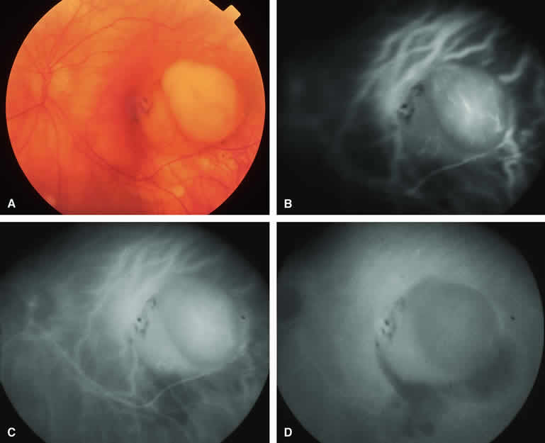
| Fig. 14. Choroidal melanoma with nodular eruption through Bruch's membrane. A. Amelanotic small macular choroidal tumor with amelanotic apical nodular eruption through Bruch's membrane and clumps of black retinal pigment epithelial (RPE) pigment along nasal margin of lesion.B-D. Indocyanine green (ICG) angiogram of lesion. B. Early-phase frame showing several prominent fluorescent intralesional blood vessels within apical nodule, generalized mild hypofluorescence of lesion, and choroidal fluorescence blockage by the RPE pigment clumps on the subfoveal portion of the lesion's base. C. Intermediate-phase frame showing increased fluorescence of tumor and reduced visibility of intralesional blood vessels. D. Late-phase frame showing greater hyperfluorescence of tumor base than of apical nodule and area of hypofluorescence along inferior margin of mass. |