|
|
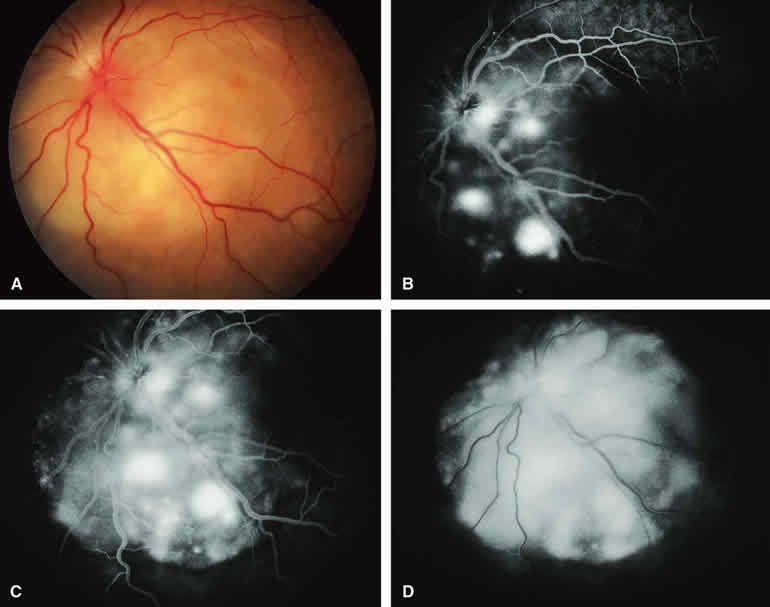
| Fig. 17. Metastatic carcinoma to choroid with prominent overlying serous subretinal fluid. A. Yellow-white juxtapapillary choroidal tumor with associated secondary retinal detachment. B-D. Fluorescein angiogram of lesion. B. Venous phase frame showing multiple intensely hyperfluorescent smudgy foci at retinal pigment epithelial level overlying ill-defined choroidal mass. C. Later venous phase frame showing generalized hyperfluorescence of mass, increased smudginess of hyperfluorescent foci overlying lesion, and abrupt demarcation between hyperfluorescent choroidal mass and adjacent normal choroid inferiorly. D. Later phase frame showing generalized hyperfluorescence of mass and overlying subretinal fluid. |