|
|
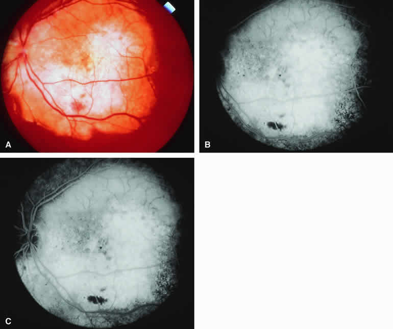
| Fig. 24. Choroidal osteoma. A. Yellowish white plate-like macular and juxtapapillary choroidal mass with well-defined margins. B, C. Fluorescein angiogram of lesion. B. Venous phase frame showing generalized hyperfluorescence of mass with spidery hypofluorescent intralesional vascular channels above and focal choroidal fluorescence blockage by blot of subretinal blood inferiorly. C. Later venous phase frame showing findings similar to those of the preceding frame. The distinct upper margin of the choroidal lesion is evident on this image. |