|
|
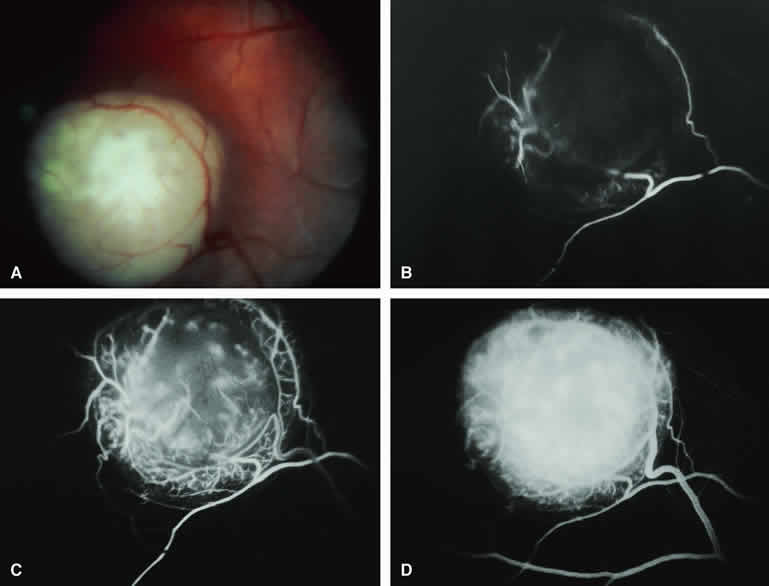
| Fig. 27. Intraretinal retinoblastoma. A. Well-defined nodular white retinal tumor with prominent retinal vasculature extending to and from lesion. B-D. Fluorescein angiogram of lesion. B. Arterial phase frame showing rapid filling of feeding retinal arteries and capillary network within lesion. C. Laminar venous phase frame showing more complete filling of tumor vasculature. D. Late-phase frame showing intense staining of entire tumor. |