
|
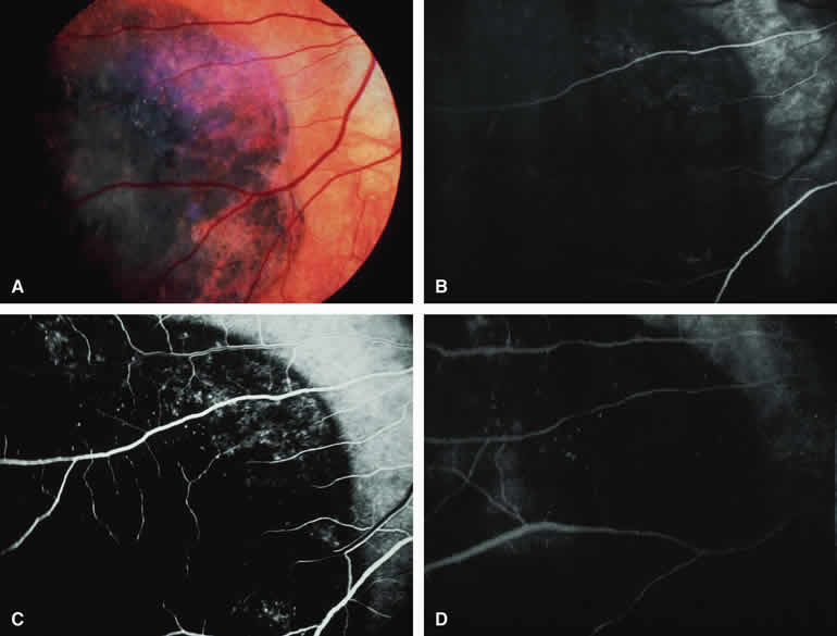
| Fig. 37. Congenital hypertrophy of retinal pigment epithelium. A. Flat gray to black retinal pigment epithelial level lesion with well-defined smooth margins. This lesion exhibits partial depigmentation peripherally. B-D. Fluorescein angiogram of lesion. B. Arterial phase frame showing hypofluorescence corresponding to lesion. Note faint transmission hyperfluorescence resulting from thinning of retinal pigment epithelium in marginal zone of lesion. C. Laminar venous phase frame showing most of lesion to be persistently hypofluorescent; however, zones of transmission hyperfluorescence are more apparent. D. Late-phase frame showing persistent hypofluorescence of lesion and fading of transmission hyperfluorescence. |