|
|
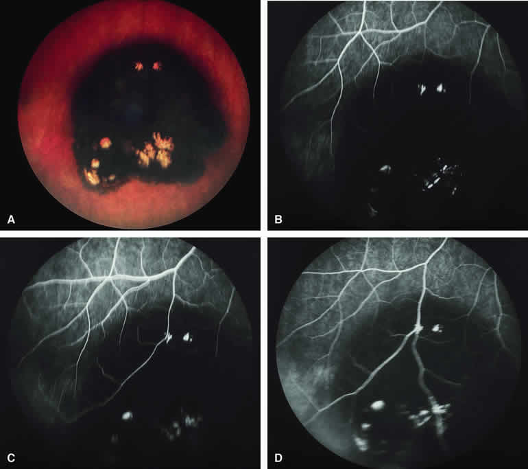
| Fig. 38. Congenital hypertrophy of retinal pigment epithelium with prominent lacunae of depigmentation. A. Well-defined black peripheral fundus lesion with several discrete foci of almost complete depigmentation (lacunae). B-D. Fluorescein angiogram of lesion. B. Venous phase frame showing well-defined choroidal fluorescence blockage corresponding to lesion and hyperfluorescent foci corresponding to depigmented lacunae. C. Later venous phase frame showing features similar to those on the prior image. D. Recirculation phase frame showing persistent hypofluorescence of lesion and sustained well-defined hyperfluorescence of focal lacunae. |