
|
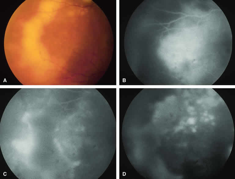
| Fig. 39. Lymphomatous subretinal pigment epithelial infiltrate. A. Geographic yellow-white subretinal pigment epithelial lesion in temporal midzone. Note smaller satellite lesions and clumped retinal pigment epithelial pigment on surface of large lesion. B-D. Fluorescein angiogram of lesion. B. Venous phase frame showing hypofluorescence corresponding to geographic fundus lesion and satellite lesions. C. Recirculation phase frame showing marginal hyperfluorescence of geographic lesion and smudgy hyperfluorescence of satellite lesions. D. Late-phase frame showing mild hyperfluorescence of geographic lesion and intense hyperfluorescence of satellite lesions. |