|
|
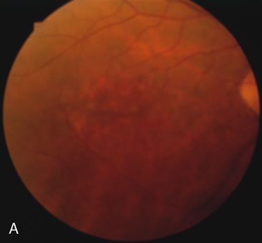
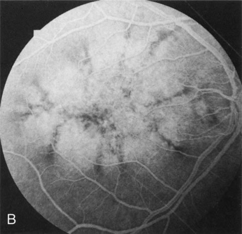
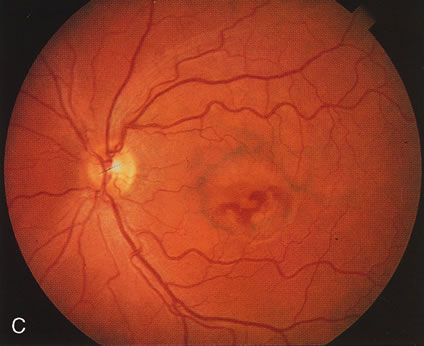
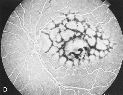
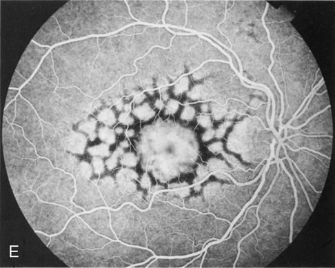
| Fig. 11. Pigment pattern dystrophies. Subtle changes of the fundus in this family member with a pattern dystrophy (A) are highlighted on angiography (B). Equally dramatic is the fluorescein angiography of this young woman who presented with poor vision in association with a retinal hemorrhage (C). The angiogram reveals a bilateral, symmetric reticular pattern of the posterior pole (D, E). |