|
|
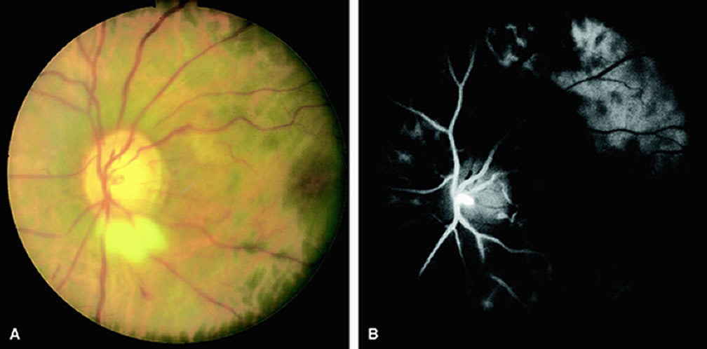
| Fig. 1. A. Fundus of a patient with the ocular ischemic syndrome. The retinal arteries appear narrowed with scattered intraretinal hemorrhages. Myelinated nerve fibers extend from the inferior margin of the disc. B. Fluorescein angiography of A demonstrates patchy choroidal filling within the peripapillary area and the presence of an abnormal leading edge of dye within a retinal artery. |