
|
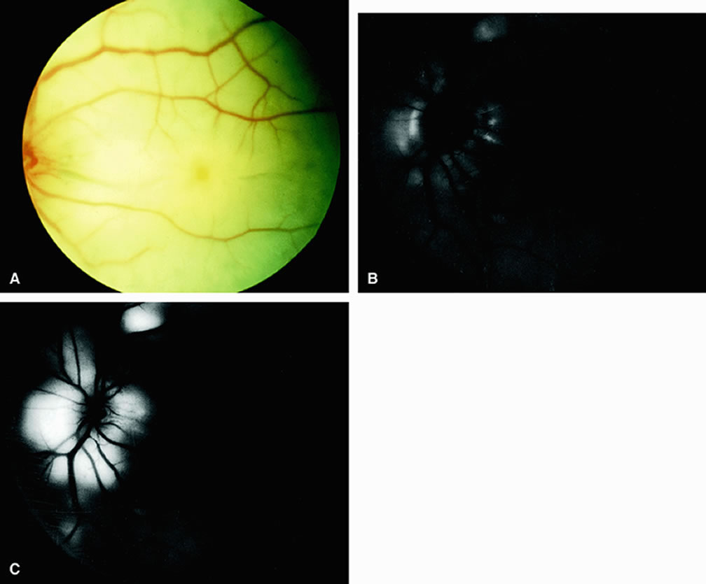
| Fig. 3. A. Acute ophthalmic artery obstruction occurring secondary to a knife injury that severed the retinal and choroidal vessels posterior to the globe. Intense retinal whitening can be seen; a cherry-red spot is absent. B. Fluorescein angiogram of A taken at 55 seconds after injection reveals an absence of dye within the retina and most of the choroid. Mild peripapillary hyperfluorescence can be seen, presumably resulting from anastomoses between the episcleral, pial, and choroidal vessels in the vicinity of the optic disc. C. Increased peripapillary hyperfluorescence at 10.5 minutes after injection. (Brown GC, Magargal LE: Sudden occlusion of the retinal and posterior ciliary circulations in a youth. Am J Ophthalmol 88:690, 1979) |