|
|
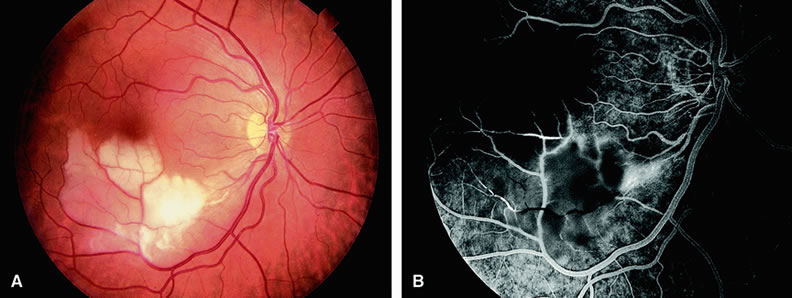
| Fig. 7. A. Patient with an inferotemporal branch retinal artery obstruction. The retina shows pallor in the distribution of the affected vessel. The other vessels appear to perfuse well. The area of pallor involves the inferior macula. B. Fluorescein angiogram of A shows retrograde filling of the affected vessel distally and retinal edema in the region of the occluded vessel. |