|
|
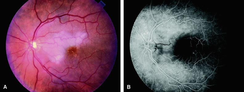
| Fig. 8. A. Fundus of an older adult patient with cilioretinal artery occlusion. The retina temporal to the optic disc corresponding to the distribution of the cilioretinal artery is edematous and white from nonperfusion. Moreover, the macula demonstrates drusen. B. Fluorescein angiogram of A demonstrates a hypofluorescent area in the distribution of the cilioretinal artery. The hypofluorescence is due to retinal artery and capillary nonperfusion. |