
|
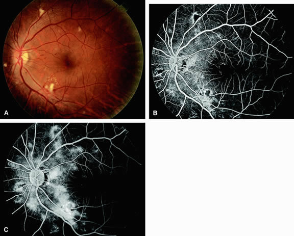
| Fig. 18. A. Fundus of a patient with radiation retinopathy. The fundus shows scattered cotton-wool spots, hard exudates, and dot-and-blot hemorrhages. Microaneurysms and macular edema are present in the region of the macula. B. Fluorescein angiogram of A shows diffuse areas of hyperfluorescence adjacent to the vascular arcades representing leakage of dye from retinal capillaries and decompensation of the retinal pigment epithelium (RPE). C. A later frame shows continued hyperfluorescence from retinal capillary leakage and more RPE leakage. |