|
|
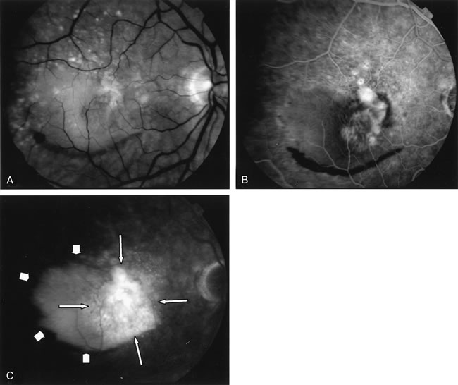
| Fig. 10. A. Red-free photograph of the right eye of a patient with wet age-related macular degeneration. There was a large, exudative pigment epithelium detachment (PED), with a narrow band of subretinal hemorrhage at its inferior border. A notch in the PED is present at its nasal edge. There were also soft drusen. B–C. Fluorescein angiography demonstrates pooling of dye into the PED (short arrows). There was also late hyperfluorescence of undefined origin consistent with occult choroidal neovascularization (long arrows). There was blockage of fluorescence at the inferior border of the PED caused by subretinal hemorrhage. |