|
|
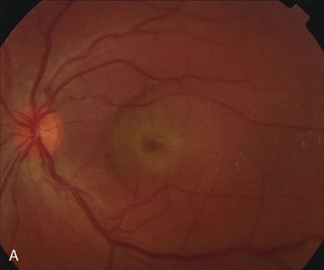
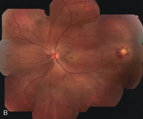
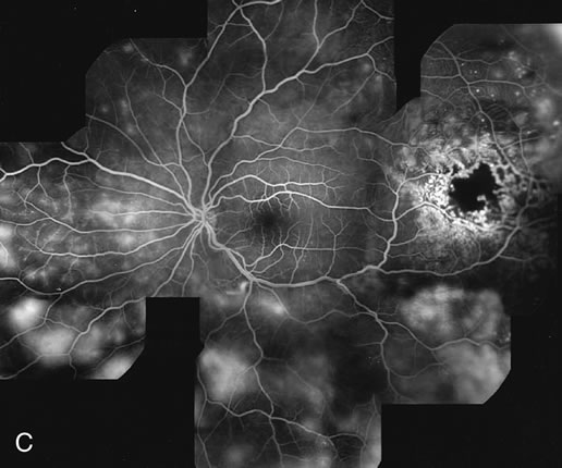
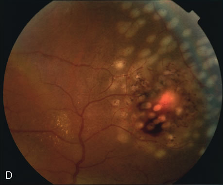
| Fig. 29. 14-year-old adolescent girl with sudden loss of vision in the left eye to 20/400. A. Clinical photograph of the posterior pole demonstrates a neurosensory macular detachment simulating central serous chorioretinopathy. B. Composite fundus photograph of the same eye demonstrates the presence of a localized area of retinal capillary telangiectasia in the temporal periphery. The intraretinal yellowish material is consistent with dehemoglobinized blood. There are also intraretinal exudates scattered throughout the fundus. C. Composite fluorescein angiogram of the same eye reveals diffuse retinal capillary telangiectasia and intraretinal leakage. The dilated retinal telangiectatic vessels are actively leaking and are responsible for the macular neurosensory detachment. This patient was diagnosed with Coats' disease. D. Scatter laser photocoagulation of the telangiectatic vessels was carried out. |