|
|
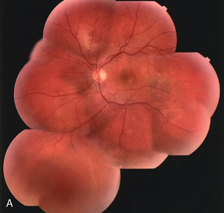
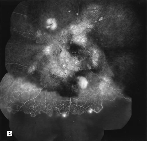
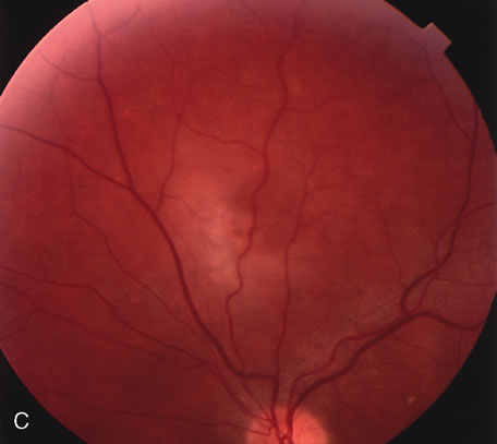
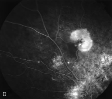
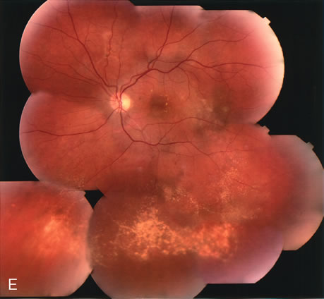
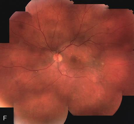
| Fig. 31. A 47-year-old woman with an18-year history of central serous chorioretinopathy in both eyes. A. Color photograph composite of the left eye shows bullous dependant detachment of the neurosensory retina inferiorly. B. Fluorescein angiogram composite reveals diffuse decompensation of the retinal pigment epithelium, multiple scattered pigment epithelium detachments 9PEDs), and obliteration of the retinal capillaries in the region of the detachments. Note the presence of early neovascularization at the junction between perfused and non-perfused retina. C. Clinical photograph of the left eye shows PED superior to the optic disc partially surrounded by fibrin deposits. D. Fluorescein angiography confirms the presence of active leakage from the serous PED. E, Color photograph composite of the same eye 2 months after laser treatment of the site of leakage reveals partial resolution of the detachment and lipid precipitation. F. Clinical photograph composite 16 months after the laser treatment in the area of the leakage shows complete resolution of the detachment and partial reperfusion of the inferior retina. |