|
|
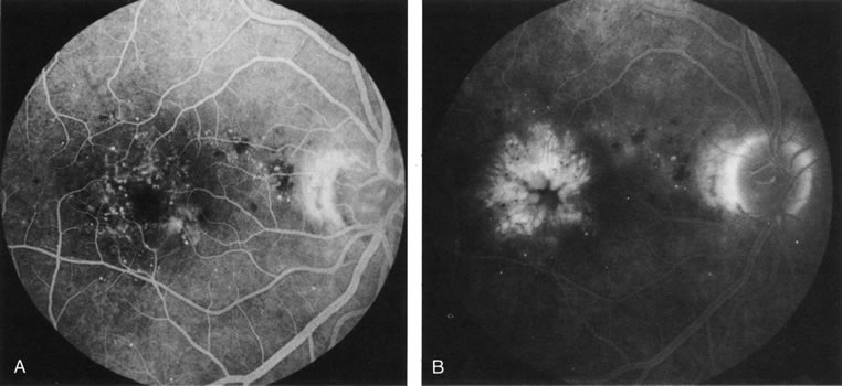
| Fig. 37. Cystoid macular edema in diabetes. A. Background diabetic retinopathy with multiple microaneurysms that hyperfluoresce and with dot and blot hemorrhages that block fluorescence. B. Late-fluorescein angiogram reveals accumulation of exudate in a cystoid pattern around the foveal avascular zone. (Courtesy of Dr. Kenneth G. Noble.) |