|
|
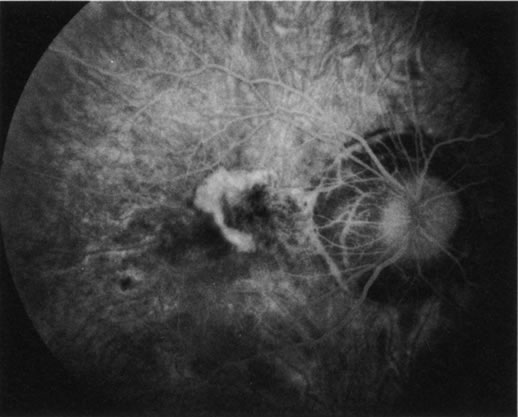
| Fig. 38. Myopic maculopathy with choroidal neovascular membrane. This patient's myopic degeneration is manifested by a large conus around the optic disc and prominence of the choroidal vasculature seen clearly through thinned retinal pigment epithelium. There is a large choroidal neovascularization (CNV) in the papillomacular area. Although CNV formation is common in myopic maculopathy, this lesion is unusually large. |