Chapter 20
ENTOPTIC IMAGERY AND AFTERIMAGES
GARY L. TRICK and ALAINA KRONENBERG
Main Menu   Table Of Contents

Search

ENTOPTIC PHENOMENA ASSOCIATED WITH PROPERTIES OF THE OCULAR MEDIA
HALOS
SHADOWS
ENTOPTIC PHENOMENA DEPENDENT ON THE STRUCTURE AND PHYSIOLOGIC PROPERTIES OF THE RETINA AND VISUAL PATHWAYS
THE EFFECT OF RETINAL PIGMENTATION
DIRECTIONAL SENSITIVITY OF PHOTORECEPTORS
ENTOPTIC VISUALIZATIONS ASSOCIATED WITH NEURAL ADAPTATION
REFERENCES

The term entoptic phenomena, derived from the Greek for “things perceived within vision,” refers to the visual sensations that arise from causes within the eye or visual system. In the most generally accepted context, the term describes the visual sensations produced or influenced by the native structures of one's own eye or visual system that are not the customary result of forming an optical image of the external environment by the refracting system of the eye. These phenomena may result from either normal anatomic components of the eye or pathologic imperfections. Although these images are not the result of normal imaging in the external world, they may be perceived as if they are produced by objects in real space. However, special methods of illumination or other types of stimulation are required to adequately create and maintain these images. In addition, the direct attention and cooperation of the observer is a critical factor for the perception of these phenomena.

Many distinguished scientists (including Aristotle, Descartes, Helmholtz, and Edison) have contributed to our understanding of entoptic phenomena. Because of their depth and breadth, Purkinje's studies are especially notable, and his name has become associated with many of the entoptic phenomena he first described.

Despite the fact that our understanding of the optical and physiologic bases of specific entoptic images remains inexact, these phenomena can be useful clinical tools. Entoptic phenomena have been used to study the pathophysiology of various ocular disorders, and they also provide a means to subjectively confirm objective findings. Entoptic imagery has been used to demonstrate the position of an opacity in the eye, measure the size of the foveal avascular zone, estimate leukocyte velocity, and the subjectively detect scotomas. However, it is important to always remember that subjective descriptions of these phenomena are notoriously inexact. Although a precise report of the visualization of a particular image can be useful, the inability to visualize an image is difficult to interpret and may simply reflect the patient's failure to understand what they are being asked to observe or their inability to accurately describe what they do perceive.

In the text that follows, the topic of entoptic imagery is subdivided into a discussion of two broad categories. One category consists of effects that are primarily dependent on the optical properties of the preretinal ocular media. The second category includes phenomena that are principally dependent on the physiology of the retina and visual pathways. It should be recognized that this classification was adopted as an aid to organization and does not imply that the two categories are independent. On the contrary, visual sensitivity and neural processing necessarily affect the perception of entoptic images in the first category, whereas the visibility of many of the phenomena in the second category is reduced when the specific optical or illumination requirements are not met.

Back to Top
ENTOPTIC PHENOMENA ASSOCIATED WITH PROPERTIES OF THE OCULAR MEDIA
Various properties of the ocular media mediate the formation of entoptic images. The vergence of light within the eye together with the optical density and refractive nature of elements in the ocular media affect the appearance of entoptic images. The translucence of an element within the ocular media plays a major role in the type of image that is formed. Similarly, the location of an element relative to both the entrance pupil of the eye and the photosensitive surface of the retina have an important effect on the appearance of an entoptic image.

The position within the eye of elements producing entoptic images can be estimated by relative entoptic parallax.1 This principle originally was described by Listing as a useful technique for demonstrating the relative position of an opacity within the eye. Essentially, moving an illuminating source (usually a point source or pinhole) affects the perceived location of the entoptic images differentially, depending on their distance from the retina (Fig. 1). If the element is behind the entrance pupil, its movement appears to be opposite to the movement of the source. Conversely, if the element is in front of the entrance pupil, it appears to move in the same direction as the source. For example, lenticular or vitreous opacities show against movement while the eyelashes show with movement. The amount of parallactic movement depends on the distance from the entrance pupil; the movement appears greater the further anterior or posterior to the entrance pupil the element is located.

Fig. 1 The use of relative entoptic parallax to estimate the position of an opacity in the eye. In the two cases illustrated here, a pinhole source is shifted downward from the optical axis (as indicated by the arrow), and the amount and direction of the displacement of the shadow of an opacity relative to the entoptic image of the pupil is evaluated. In both cases (A and B), the shadow of the opacity (black shadow) appears centered within the entoptic image of the pupil (red lines) when the pinhole is positioned on the optic axis. In the first case (A), the opacity is positioned posterior to the entrance pupil of the eye. When the pinhole is shifted downward, both the entoptic image of the pupil (green lines) and the shadow of the opacity (yellow shadow) are displaced upward, but there is relatively less displacement of the shadow. As a result, the shadow of the opacity is no longer centered in the entoptic image of the pupil. This is perceived as if the shadow moved in the opposite direction from the image of the pupil. The closer the opacity lies to the retina, the greater the relative displacement. In the second case (B), the opacity is positioned anterior to the entrance pupil of the eye. Once again when the pinhole is shifted downward, both the entoptic image of the pupil (green lines) and the shadow of the opacity (yellow shadow) are displaced upward, but in this case there is relatively more displacement of the shadow. As a result, the shadow of the opacity is no longer centered in the entoptic image of the pupil. This is perceived as if the shadow moved in the same direction as the image of the pupil. The closer the opacity lies to the pinhole, the greater the relative displacement.

A more precise method for measuring relative entoptic parallax was developed by Brewster and modified by Donders (Brewster-Donders method for measuring the depth of opacity). In this method, a Scheiner disc with two small pinholes separated by approximately 2 mm. is positioned in the anterior focal plane of the eye. The field of view is then bounded by two overlapping circles whose width depends on the diameter of the exit pupil of the eye. Because the pinholes serve as two distinct sources, an opacity will cast two shadows on the retina. The distance between the two shadows, when projected onto a screen in real space, can be determined. As long as fixation is constant, this distance can be used to accurately estimate the location of the opacity.

Back to Top
HALOS
Because of irregular refractivity, translucent elements scatter transmitted light, reducing image contrast and forming halos. When the translucent element is more refractive than the surrounding ocular media, the entoptic image is relatively bright and surrounded by a dark rim (diffraction ring). A translucent element that is less refractive than its surrounding media produces a dark image surrounded by a light rim. If the translucent element is large or located close to the retina, a true scotoma results.

Ocular structures that act as diffraction gratings produce diffraction spectra when a point source is viewed. Entoptically, these diffraction spectra are visible as rainbow-like halos or coronas centered on the source. With a white light source, the outside rings of the diffraction spectra are red, while the inner rings are blue–violet. Halos are caused by diffraction from small regularly sized or spaced structures within the eye. The specific characteristics of individual halos depend on the structure of the diffraction grating underlying their generation. Diffraction theory predicts that the angular diameter of a halo is related directly to the wavelength of the light and inversely to the size (or in the case of a regular pattern to the spacing) of the diffracting structure. Therefore, as the diameter of the diffracting structures increases (e.g., cells, droplets) or the spacing of the elements in the diffraction grating gets larger (e.g., lens fibers) the diameter of the halo will become smaller. The longer the wavelength of light, the larger the diameter of the halo. The perceived diameter of an entoptic halo also depends on the distance between the diffracting structures and the retina. Halos are smaller when the diffracting structures are nearer to the retina.

Halos can result from either the normal physical properties of ocular structures or the alterations in these physical properties caused by the response of ocular tissues to pathology. In the healthy eye there are differences in size between halos associated with different ocular structures.2,3 With the exception of the zonular insertions and anterior cellular layer, the crystalline lens is composed of fibers that pass in an approximately radial manner from the anterior to the posterior sutures. As a result, the crystalline lens may be regarded as a strong positive lens with a radial diffraction grating (similar to any two- or three-dimensional array of lines or dots showing periodic variations of either transparency or refractive index) superimposed on the periphery. Because the axial portion of the lens appears to be relatively uniform, no lenticular halo is observed with a pupil less than 3 mm in diameter. With a dilated pupil, the effect of the peripheral lens becomes marked. In this case, the halo that generally is most visible results from diffraction by the radially arranged lens fibers. This produces a halo with diffraction rings that have been estimated to have diameters ranging from 4.5 degrees for violet, 5 degrees for blue, 5.4 degrees for green, and 6 degrees for yellow.4 A larger halo (approximately 9 degrees) was first described by Descartes in 1637 and appears to be caused by second-order diffraction by the corneal endothelium. A smaller halo (diameter of approximately 3 degrees) is a result of the structure and organization of the corneal epithelial cells, whereas the corneal endothelial cells and/or lens epithelial cells contribute to a slightly larger halo (approximately 4.5 degrees in diameter).

The halos associated with the response of ocular tissues to disease conditions include a halo of 7 degrees to 12 degrees that results from edema of the deeper layers of the corneal epithelium such as might be associated with elevated intraocular pressure (IOP), aphakic bullous keratopathy, and ultraviolet keratopathy. A larger (12 degrees to 14 degrees) halo is produced by debris such as mucus, blood, pus, or particulate matter on the corneal surface In some cases, the halo resulting from corneal edema can be differentiated from the lenticular halos by placing a stenopeic slit before the eye and restricting light entering the eye to a small zone of the entrance pupil (Fig. 2). Passage of light through a particular section of the lens generates a specific double section of lenticular halo. Moving a vertical stenopeic slit across the pupil gives moving sectors of light corresponding to the portions of the lenticular halo that are eclipsed (partly or wholly) according to the area of pupil that is occluded. Conversely, when the corneal stroma or epithelium becomes edematous, the regularity or spacing of the fibrils is disturbed, leading to small-body diffraction, with each element scattering light equally in all directions. As a result, the halo produced by corneal edema only appears reduced in intensity when a stenopeic slit passes in front of the eye.

Fig. 2 When a narrow (1 mm or less) stenopeic slit is positioned in front of the pupil, a lenticular halo is visualized (lines A and B). The visible portion of the halo formed (line B) depends on the orientation of the lens fibers in the regions of the pupil that are illuminated (line A). When the stenopeic slit is moved sequentially across the pupil (C), a lenticular halo that resembles the rotating arms on a windmill will become evident (D).

Back to Top
SHADOWS
Nontranslucent structures absorb light and are opaque. Consequently, nontranslucent structures cast shadows. If the nontranslucent element is nearly the same size as the pupil or is situated close to the retina, these shadows are detectable. Otherwise, the shadow is not detectable because only the diffraction at the edges of nontranslucent elements produces light scatter. Therefore, the visual problems associated with nontranslucent elements are most severe when they are located posteriorly and produce a positive scotoma.

The pupil, lids, lashes, tear film, and corneal mucus can be observed entoptically as shadows. Under optimum conditions of illumination, the entoptic field is bounded by the shadow of the inner edge of the iris, which is visible as a bright patch limited by a circular ring with ragged edges corresponding to the pupillary margins. Irregularities of the pupillary margins as well as contraction and dilatation can be detected in this image, which also can be used to monitor the consensual pupil reaction driven by changes in illumination in the contralateral eye. Cogan5 used the entoptic visualization of the pupillary margins as the basis for a simple technique that permits patients to measure the size of their own pupil. When a pinhole is held close to the eye and a uniform field is viewed, the field appears to be the same size as the pupil. If two separate pinholes are viewed simultaneously, the patient sees two images the same size as the pupil. When the two spots appear juxtaposed, the distance between their centers equals the diameter of the pupil.

Superimposed on the bright image of the pupil, the shadows of the lashes appear to be jumping in and out of view when the lids are moved. In this image the lower lashes appear uppermost because of image inversion by the optics of the eye. The tear film and the mucus on the corneal surface also produce entoptic images. Horizontal striations that change with blinking typically result from visualization of the tear film. This can be demonstrated by partially closing and then opening the eyes while viewing a uniform field. A bright longitudinal stripe, with a dark margin caused by a ridge of tears, should be evident. Corneal mucus, as might result from inflammation, produces shadows that appear as bright spots surrounded by dark rings that move up and down as the palpebral fissure is narrowed or widened. Because mucus is very adherent to the cornea, these shadows can be quite persistent.

Folds in corneal epithelium create shadows that also appear as horizontal bands of light. These shadows are visible as unbroken lines extending across the entire width of the pupillary image, and they change their position as the eyelids are slowly opened or closed. Shallow linear channels in the corneal epithelium, such as those created by gentle digital pressure on the cornea through the closed eyelid or by rigid contact lens wear, also can be visualized entoptically. These shadows typically appear as mosaic patterns and are believed to be produced by elevated ridges in Bowman's membrane that are created during the period of corneal flattening.

Structures in the lens and vitreous also cast shadows. The poor focusing of marginal rays through the pupil, caused by fibrous composition of the lens, produces images that appear as a system of rays that can be observed emanating from small bright objects on a dark background. A typical example is a star that appears to have a spiky surround. If the pupil is replaced by a smaller adjustable diaphragm, the rays (spikes) expand and contract with variations in the diaphragm. In cases in which the nucleus of the lens has a significantly different focus from the periphery, the central bright image may appear broken into multiple images (polyplopia). In addition, opacities in the lens and lenticular cataracts may be evident as dark, granular, stable patterns if their shadows extend to the retina. Alternatively, lenticular opacities may reduce retinal illumination and scatter light without producing detectable shadows.

Small inclusions in the vitreous (vitreous floater) may be visible entoptically as mobile shadows resembling bubbles, strings of pearls, or bundles of filaments. With a point source of white light, colored fringes may be detected bordering these shadows. Vitreous floaters can change shape when the eye moves. One form of vitreous floater casts a delicate, somewhat lacy or chain-like shadow that moves with gaze but tends to overshoot fixation and then return when the eye movement stops. Floaters in the anterior vitreous are perceived to move in a direction opposite to the movement of the eye, whereas floaters in the posterior vitreous move in the same direction as the eye movement. These entoptic visualizations often can be seen without the aid of a pinhole because they are caused by fine opacities located near the retina. On the basis of their appearance, these floaters have come to be known as muscae volitantes (Latin for flying gnats). Vitreous floaters are common, frequently visualized in the normal eye, tend to increase with increases in myopia or age and often are a source of concern to the patient. Once developed, a vitreous floater may persist for life, but the sudden onset of numerous floaters may be the initial sign of a retinal detachment. The vitreous floater associated with retinal detachment may represent blood particles in the vitreous. In cases in which these floaters disappear after a few days, it should not be assumed that the underlying problem has resolved.

Back to Top
ENTOPTIC PHENOMENA DEPENDENT ON THE STRUCTURE AND PHYSIOLOGIC PROPERTIES OF THE RETINA AND VISUAL PATHWAYS

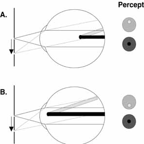
IMAGES CAUSED BY THE RETINAL CIRCULATION

One of the most easily demonstrated of the entoptic visualizations is the subjective perception of the retinal blood vessels. Under ordinary circumstances, the shadows cast by the retinal vessels remain in fixed position relative to photoreceptors, even during eye movements. Because the underlying neural network is adapted, the vessels are not subjectively apparent in this situation. However, Purkinje noted that when light enters the eye from an unusual angle (e.g., obliquely through cornea or through the sclera) the shadows of the retinal vessels fall on unadapted retina and the shadows of the retinal vessels become visible for a brief period. This can occur, for example, during slit lamp biomicroscopy of the anterior segment. The shadows of the arterial and venous tree will be perceived as a greatly magnified, inverted image that has come to be known as either Purkinje's tree or the Purkinje figure. In this image the retinal vessels are observed arising from the optic nerve head and branching out with finer and finer branches extending toward the fovea. With this type of viewing, the foveal pit, which lacks blood vessels, is readily distinguishable in this image as a vacant area surrounded by the branching pattern. Helmholtz made use of the parallax intrinsic to this phenomenon to demonstrate that the light-sensitive elements in the retina lay within the 
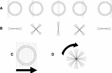
photoreceptor layer.

To visualize the retinal vessels, place a small, bright light source over closed eyelids near the limbal margins and oscillate the light source at a relatively low frequency (2 to 4 Hz). In the resulting entoptic image, the retinal vessels will appear as dark shadows against a yellow–orange background. To be able to perceive greater details in the image (such as the retinal capillaries), gaze at a uniformly illuminated, bright surface through a pinhole positioned at the anterior focal plane of the eye. The parallel light rays obtained with this technique enhance the contrast of the image so that shadows of vessels, including the capillaries surrounding the fovea, become visible. With sustained viewing, the Purkinje figure will fade, but the image can be regenerated by simply repositioning the illuminating source. By rotating the light source in a circular pattern, image stabilization can be avoided, and the shadows of the vessels will be perceived continuously.

Visualization of the retinal capillaries surrounding the fovea permits estimation of the size and shape of the foveal avascular zone. Applegate and colleagues6,7 used entoptic visualization of the retinal capillary net to measure the size and shape of the foveal avascular zone. They determined that the foveal avascular zone is an elliptical region with a mean diameter of approximately 0.755 mm and a major axis approximately 17% longer than the minor axis.7 These dimensions are consistent with foveal avascular zone diameters measured by angiographic techniques.8,9 Zeffren and co-workers10 also found that the point of fixation often is not centered in the foveal avascular zone. Furthermore, they observed that in a number of cases the point of fixation is located eccentric enough within the region that macular photocoagulation to within 200 μm of the center of the region could result in a lesion at the point of fixation, thereby compromising visual acuity.

With conscious effort, pulsation of retinal blood vessels can be seen after physical exertion or pressure on the globe. On close observation, it is evident that: (1) the pulsation proceeds recurrently along certain pathways; (2) the shadows are not seen in an area close to the point of fixation; and (3) the movement is not uniform but rhythmic or pulsatile. Two quick changes in the appearance of the vascular network surrounding the fovea can become evident. In one phase there is a rapid expansion of arterial tree that is synchronous with cardiac systole and corresponds to the steep and rapidly moving ascending limb of the pulse. This phase is immediately followed by a slower contraction that corresponds to the descending limb of the pulse wave. Because the period of the contraction is of longer duration than the period of the expansion, the contraction often appears more prominent.

If accommodation is relaxed and a brightly illuminated uniform blue–violet (350 to 450 nm) surface (e.g., blue sky or blue monitor) is viewed, then the circulation within the retinal capillaries also can be visualized entoptically. Darting points or spots that appear as bright circles with dark centers against a relatively darker background will be visible. The darting points may appear to have short tails and to follow sinusoidal paths similar in shape to the retinal capillary loops evident on trypsin digest preparation. By viewing the spots with one eye and the Purkinje tree with the other eye, Marshall11 found the spots to be on one plane and the superficial retinal vessels on another plane. Therefore, he concluded that the spots were deeper than superficial retinal vessels, possibly representing cells within the deep capillary bed at the level of the inner plexiform and inner nuclear layers. Considerable additional evidence has been accumulated to suggest that this entoptic visualization results from the movement of blood cells. This includes evidence that these darting points cannot be seen in the central fovea and that pressure on the eye slows their movement. In fact, with pressures in excess of 50 mm Hg, the movement is halted. Although the type of blood cells responsible for this phenomenon is still in question, evidence strongly favors leukocytes, because the points are too infrequent to be erythrocytes. This could be explained by assuming that erythrocytes are only visible when passing through deep capillary bed. Hemoglobin, however, has an absorption peak in the blue–violet range, so erythrocytes would cast relatively intense, continuous shadows against a light background. Under the same conditions, leukocytes would transmit blue–violet light and would be seen, therefore, as bright interruptions in a continuous shadow. This latter description is consistent with the subjective report of the appearance of this entoptic image.

Riva and Petrig12 developed a technique that uses the properties of this entoptic image to estimate leukocyte velocity. Patients are asked to match the velocity of global motion in a simulated pattern of random dots to the global motion of the darting points in the entoptic image from their own eye. The density of points (shadows) in the simulation, the time average of the velocity waveform, and the pulsatility of the motion are adjusted to match the patient's own entoptic visualization. Shadow velocity is then used to estimate volume blood flow. The effects of hyperoxia and hypoxia, changes in perfusion pressure, cigarette smoking, and diseases affecting the visual system (e.g., diabetes and glaucoma) on retinal blood flow have been studied using this technique.13

With further intense concentration, the perception of these luminous darting points may be observed to be followed by the visualization of a darkened field that has the appearance of a turbulent surface of boiling water with an abundance of smaller bright points moving much faster than the points detected in the earlier image. Distinct islands of boiling can be appreciated within the turbulent surface. Marshall11 describes this image as “A surging circulation in irregular sinuses somewhat fanlike in appearance of a dark reddish-gray color bounded by a black background meshwork. Over the field may also be seen innumerable fine granules of black…but not always. The effect lasts only a few seconds, but it may usually be repeated several times.” One possible explanation of the source of this perception is that it represents a visualization of circulation in the choriocapillaris.

PHOSPHENES

The term phosphenes (Greek for “to show light”) refers to the visual sensations of light produced by nonphotic or nonluminant stimuli. Mechanical and electrical energy as well as electromagnetic energy outside the visible spectrum may be the source of different phosphenes. These forms of energy often are termed “inadequate” stimuli because, under most circumstances, they do not produce visual perception.

Various forms of mechanical energy produce phosphenes. Pressing or rubbing the eye, blunt trauma, strenuous exercise, sudden eye movements, and prolonged accommodation or convergence all are known to bring about phosphenes. Gentle, but forceful pressure to the nasal or temporal portion of the eye elicits an immediate, very bright, well-circumscribed ring of blue–white in the periphery of the opposite visual field (Purkinje's blue ring). This is best seen in a dark-adapted eye and has been attributed to a mechanical distortion of the sensory elements in the retina, which then initiates a neural response. When pressure on the globe is maintained for a prolonged period (approximately 3 minutes) at a level that is sufficient to create slight discomfort, a perception of a broad circular blue ring often can be detected. This percept has been described as a broad automobile tire somewhat flattened vertically. On careful observation, this image may initially be observed to consist of two blue spots (one nasal and one temporal) that expand slowly to take the form of a broad arc. As the figure continues to grow, the arcs coalesce into the blue circle by a slow motion (similar to water seeping between two plates of glass). The center of the oval, which is devoid of color, extends from approximately 2 degrees above and below the fovea to approximately 3 degrees on either side. The periphery of the oval is sharply outlined and extends to approximately 10 degrees vertically and 12 degrees horizontally. As a result, this image approximates the outer limits of the macula, with the blue circle covering the retinal region of greatest rod density.

Several different phosphenes are associated with eye movements. If an eye movement is made while staring at an evenly illuminated bright surface, a dark gray or pale blue oval area ringed by a bright blue–white border may be visualized in the region of the blind spot of each eye (the fiery rings of Purkinje). The rings appear accentuated in the dark and often are more evident in the eye moving nasally. Generally, the rings are perceived as larger than the blind spot.1 One possible cause of this phosphene is pressure on the retina adjacent to the optic disk caused by traction from the optic nerve. During nasal rotation, traction is exerted on the retina temporally adjacent to the disk while the adjacent area on the nasal side is under compression. More extensive eye movements produce similar phosphenes that are observed farther in the periphery and may result from traction on the retina by the extraocular muscles at their points of insertion adjacent to the ora serrata.

When dark-adapted, a different phosphene (Nebel's flick phosphene) may be observed after a rapid flick or microsaccade.14 This phosphene typically is evident in people older than age 40 years and is perceived as a brief (0.33 seconds), bright, blue or orange, sheaf-like pattern extending 20 degrees to 40 degrees horizontally and vertically and centered on a dark background. With repeated observation, the color fades from this phosphene and the pattern may shrink. The apex of the pattern points in the direction of the eye movement and, similar to Purkinje's fiery rings, localizes at the blind spot. Typically, this pattern is observed in both eyes, although the pattern may be larger and brighter in the eye moving nasally. Nebel14 proposed that this phosphene is caused by a transient deformation of the posterior vitreous face, perhaps because of early posterior vitreous degeneration. In an early stage of posterior vitreous degeneration, there may be a slight looseness and shrinkage of the posterior vitreous, decreasing the normal slack and lag while increasing the force to which the retina and vitreous attachments are subjected. Abrupt eye movements transmit the inertial drag of the vitreous body to the retina at the posterior pole (centered on the optic disk), causing deformation of retinal structures similar to a mechanical phosphene.

Degeneration of the vitreous with frank posterior detachment appears to underlie the entoptic visualization known as Moore's lightning streaks. Mechanically related to, but distinct from, Nebel's flick phosphene, Moore's lightning streaks appear as flashes of light that often are described as looking like lightning. Moore described them as having a vertical direction and only occurring on the temporal side of the eye.15 Verhoeff,16 however, found that they could occur in both the nasal and the temporal visual fields. Verhoeff theorized that this entoptic phenomenon resulted from degenerative condensation and vitreous shrinkage with separation from the retina. The lightning streaks are generated when an eye movement causes a sudden impact of condensed vitreous on the retinal periphery. Vitreous traction on the retina was excluded by the fact that the streaks appear on the side to which the ocular rotation is directed. Moore's lightning streaks occur in association with the development of opacities in the vitreous, seldom occur before middle age, and are more frequent in females. Moore suggested that although the streaks may persist for years (possibly becoming less frequent and less brilliant with age), they are not precursors of retinal detachment and they do not imply or herald any serous disease of the eye. In an extended follow-up (18 years or more) of patients reporting Moore's lightning streaks, Verhoeff16 was unable to find any patients returning with retinal detachments in the affected eyes. In addition, Linder17 found that 35% of a series of patients (n = 115) with posterior vitreous separation observed the lightning streaks; however, none of these had a retinal detachment. These reports support the belief that lightning streaks are innocuous. Nevertheless, individuals reporting light flashes should receive careful and periodic examination of the retina and vitreous to rule out the existence of significant peripheral retinal degeneration or retinal detachment. In this regard, it is important to differentiate the lightning streaks from nonspecific flashes, which may indicate the presence of vitreoretinal pathology. Berens18 found that 7 of 36 patients reporting nonspecific light flashes had posterior vitreous separation and rhegmatogenous retinal detachment, whereas Morse and co-workers19 found that 23 of 100 patients reporting nonspecific light flashes had vitreoretinal disease (16 with retinal breaks or holes).

A sudden accommodative response also can produce a phosphene.20 This entoptic image appears similar to the image produced by a more forceful eye rotation, but in this case the cause is most likely to be traction of the ciliary muscle on the peripheral retina. This explanation is consistent with evidence that the ora serrata moves forward during accommodation (0.5 mm per diopter of accommodation). During overaccommodation when viewing an object close to the limit of accommodation, the whole field tends to darken somewhat nonuniformly. The periphery is affected more, but a central patch is also dark. On further accommodation, the central patch lightens in its center. No loss of acuity is evident in the darkened areas. This phosphene can be appreciated through an artificial pupil and the patchiness is not consistent with the more general reduction in luminance associated with pupillary constriction.

Electrical stimulation of the eye also produces phosphenes.21–22 When the terminals of a low-voltage cell (less than 10 volts) are placed between the tongue and the upper lip while in the dark, a faint glow can be observed all over the visual field. These phosphenes have been described as being very distinctive and conspicuous, appearing almost as sharply delineated as real objects. The phosphenes also are attractive, similar to contour lines on topographic maps except that the lines do not cross one another and they disappear either by moving off into the periphery or by forming a loop that eventually contracts to nothing. With alternating current, seven types of pattern may be observed. Four of the patterns are noted when the eye is uniformly illuminated, whereas the other three are detected in the dark-adapted eye. These patterns also appear to be dependent on the frequency and the current of AC stimulation. Wolff and colleagues21 suggested that the patterns arise in the retina as a result of the alternating current acting on radially oriented structures such as the photoreceptors and bipolar cells. Carpenter21–22 further suggested that different types of patterns arise within neuronal domains responding to opposite phases of the current.

Exposing the eye to electromagnetic sources such as x-rays, cosmic rays, and fast particles that are outside the visible spectrum also causes phosphenes. The phosphene associated with exposure of the eye to x-rays (100 to 0.01 nm) appears as a homogeneous luminous blue–green or yellow–green glow that fills the entire visual field and resembles an atmospheric electrical discharge behind clouds on the horizon. The phosphene does not appear to be caused by fluorescence of the retina or ocular media. Rather, it is thought that x-rays have a direct ionizing effect on the rod and cone photopigments. Because this phosphene is only perceived in eyes with light perception, it has been used to test retinal function in eyes with opaque media. In contrast to the phosphene elicited by exposure to x-rays, the phosphene associated with radium exposure is a diffuse homogeneous glow that is more green than blue. The radium phosphene is primarily the result of beta particles acting on the ocular media to induce fluorescence, although a lesser portion of the effect may be caused by gamma rays acting in a fashion similar to x-rays.

During space flight, astronauts have reported entoptic visualizations, many of which only occur in the dark-adapted eye. These entoptic phenomena have been described as colorless or blue-white light flashes and streaks of light. The mechanism mediating these effects is uncertain. However, based on evidence that cosmic rays can be detected by the human eye,24 it has been speculated that cosmic particles that penetrate the space capsule play a role in these visualizations. An alternative explanation is that these phosphenes are caused by radiation that is produced whenever a charged particle passes through a transparent medium (e.g., vitreous or retina) at a speed exceeding the speed of light in the same medium (Cerenkov radiation). This produces shock-wave phenomena (the optical equivalent of the sonic boom) or direct ionization with excitation of retinal molecules secondary to high-energy proton recoils together with the release of alpha particles by neutron reactions on carbon, nitrogen, and oxygen molecules. This alternative is supported by evidence that neutrons, protons, alpha particles, muons, and the nuclei of carbon, nitrogen, and hydrogen cause similar sensations in ground-based simulations. The visible sensations associated with Cerenkov radiation are only apparent in the dark-adapted eye and include large crescent-shaped and cloud-like flashes, brighter and smaller flashes, and wide streaks or bands with dark centers. The visible sensations associated with direct ionization and excitation include pinpoint and star-like flashes and short light streaks.

BLUE ARCS OF THE RETINA (PURKINJE)

During monocular viewing of a dim light of any color in a darkened room with the temporal parafoveal retina stimulated (i.e., 1 degree to 2 degrees from the fixation point), two faint glowing blue–gray arcs will be seen bowing above and below fixation (Fig. 3). These arcs begin at the source (although they are noticeably wider than the source) and extend to the blind spot. If the nasal parafovea is stimulated instead, then a triangular patch of blue haze (a blue spike) with its base at the light source and its apex at the blind spot will be observed. This entoptic phenomenon, which is referred to as the blue arcs of the retina, appears briefly (0.5 to 1 second) but fades on extended observation. The position and orientation of the blue arcs are generally held to correspond to the route of the parafoveal arcuate nerve fiber bundles extending to the optic disk. Thus, this visualization is thought to be the result of secondary electrical stimulation of the retina whereby action potentials in the arcuate bundles excite adjacent neurons. Moreland25,26 suggested that both rod and cone pathways are involved in generating this entoptic image.

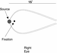
Fig. 3 An illustration of the appearance of the blue arcs of the retina when the temporal parafoveal retina is stimulated.

POLARIZED LIGHT

The eye's response to polarized light is evident in the entoptic phenomena known as Haidinger's brushes. These entoptic images appear as faint yellow and blue brush-like patterns in the central visual field when a source of polarized light is viewed (Fig. 4). The images extend from the fixation point in a pattern similar to a Maltese cross or a windmill, with the yellow brushes standing out against a blue background when white light is used. Against a blue background, the blue brushes may look black. The blue arms coincide with the electric vector of the polarized light, whereas the yellow brushes are oriented perpendicular to the polarity of the source. Although this entoptic phenomenon may be seen in the naturally polarized light of the sky it is more readily demonstrated by looking at a bright, diffuse white or blue field through a sheet of polarizing material that is continuously rotating. The rotation helps to prevent adaptation so that the effect persists. If the rotation of the polarizer is halted, the visualization fades rapidly.

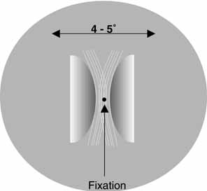
Fig. 4 An illustration of the appearance of Haidinger's brushes in the central visual field when a source of polarized light is viewed. The images extend from the fixation point in a pattern similar to a Maltese cross or a windmill, with the yellow brushes standing out against a blue background when white light is used. The blue arms coincide with the electric vector of the polarized light, while the yellow brushes are oriented perpendicular to the polarity of the source.

Haidinger's brushes apparently are caused by the fact that some structure of the eye acts as a radial analyzer for blue light. In 1844 Van Haidinger described the brushes as an entoptic phenomenon caused by the birefringent properties of the ocular media. Helmholtz assumed that the yellow pigment overlying the photoreceptors within the macula was birefringent and could act as a polarizer. This yellow screening pigment is found within a region of 6 degrees to 10 degrees from the fovea (an area called macula lutea), and Wald found this pigment to be the carotenoid xanthophyll with an absorption maximum at 430 to 490 nm. Attempts to quantify the absorption of blue light by this yellow pigment have been made by measuring the visibility of Haidinger's brushes at different wavelengths. DeVries and colleagues27 and Naylor and Stanworth28 both demonstrated that the spectral distribution of the effect was virtually indistinguishable from the optical density spectrum of the macular pigment. Although the origin of macular dichroism has not been fully explained, the radial structure of the Henle fiber layer appears to play an important role. Bone29 and Bone and Landrum30 suggested that the radially arranged nerve fibers between the inner and outer limiting membrane of the retina provide a matrix for the alignment of xanthophyll molecules. Alternatively, Hemenger31 has argued that form dichroism (arising only from the radial structure of Henle's fiber layer and not from the preferential orientation of molecules) must contribute to the phenomena. Additionally, the cornea and lens may have an effect on the polarization of the incoming light. In particular, the birefringent properties of the collagen fibrils within the cornea could influence the orientation of the brushes, because the collagen in the corneal stroma is predominantly oriented in an upward and outward diagonal.32 This contribution from the preretinal media could explain why patients with keratoconus have difficulty perceiving this entoptic phenomenon.

The visualization of Haidinger's brushes has been applied in several different clinical contexts. Because the effect is caused by the orientation of elements in front of the photoreceptors, any process disrupting this orientation without severely affecting the photoreceptors or retinal circuitry could lead to a reduction in the visibility of the brushes while minimally affecting visual acuity. For example, patients with central serous retinopathy or macular edema may be unable to detect the brushes at visual acuity levels (20/40 to 20/80) that exceed the level to which acuity must be reduced by lenticular opacification to obscure the effect (20/200 to 20/400). The visualization of Haidinger's brushes also has been used to determine the angle between fixation and foveal axes in amblyopic eyes with eccentric fixation. In addition, the phenomenon has been used in the diagnosis and treatment of binocular suppression scotomata associated with some forms of esotropia.

Back to Top
THE EFFECT OF RETINAL PIGMENTATION
The filtering effect of the xanthophyll in the central fovea contributes to another entoptic visualization, Maxwell's spot. In this case, the shadow of the macular pigment is detectable when equally bright green and blue light are flickered in counterphase. Bringing this light to a narrow focus in the pupillary plane reveals a very fine, discrete mottling in the center of the visual field with a vague dark shadow at the point of fixation. Maxwell's spot also can be observed by suddenly interposing a blue filter in front of a brightly illuminated uniform field. The shadow extends approximately 1.5 degrees above and below fixation and is slightly elongated horizontally. Under optimum conditions, the spot may be resolved into distinct, irregular rings (ragged bulls-eye). Often, the shadow appears to be surrounded by a blue halo.
Back to Top
DIRECTIONAL SENSITIVITY OF PHOTORECEPTORS
The Stiles-Crawford effect demonstrates the directional sensitivity of the retinal photoreceptors.33 Parallel rays entering the pupil near its center are more effective in stimulating retinal cones (and to a lesser extent rods) than rays that enter more peripherally. The effect is symmetric (falling off evenly with distance from pupil center) except in eyes that are not normal, where an asymmetry may be evident. This effect appears to be the result of the manner by which the photoreceptors collect and channel incident light, acting like waveguides or fiberoptics. Consequently, the Stiles-Crawford effect can be used to evaluate photoreceptor alignment or misalignment in retinal disease.34
Back to Top
ENTOPTIC VISUALIZATIONS ASSOCIATED WITH NEURAL ADAPTATION
Complete dark adaptation of the eye does not produce a sensation of absolute black. Instead, a uniform gray with superimposed phosphene-like dots is generally noted. The dots observed in this entoptic phenomena are more mobile than phosphenes and may form colored patterns that drift about the field. This effect has been termed “dark light,” retinal “self-light,” or the Eigengrau. Shifting patterns of spontaneously occurring neural activity in the retina and afferent pathways resulting from thermal isomerization of photopigment molecules, random activity of visual neurons, and indigenous neurotransmitter release appear to be the source of Eigengrau. Some subjects report more complex patterns in the Eigengrau that may be associated with spontaneous neural activity leading to coupled activation of neurons in higher visual centers.35

Another example of the influence of neural adaptation on entoptic visualization is the Troxler effect. Troxler fading refers to the spontaneous suppression of the visibility of an image that occurs when one stares intently at a point in a scene. After some seconds or even minutes, parts (but not all) of the field will lose contrast and merge into a misty blur. Moving the eyes restores clear vision. This is often reported by patients who maintain very steady fixation while receiving a pattern-reversal visual-evoked potential. A similar effect occurs when an image is stabilized on the retina with the eyes free to move. In this case, however, there is a complete loss of the contrast of the image.

This entoptic phenomena has become the basis for a unique form of subjective perimetry, which has been shown to be effective in detecting peripheral retinal lesions. Peripheral scotomas are normally repressed by the visual system because of the Troxler phenomenon. This makes it difficult for a patient to detect a scotoma in their peripheral visual field. However, entoptic perimetry overcomes this difficulty. Entoptic perimetry is performed using a television with random particle motion on a black and white (“snow field”) background. The patient is asked to fixate in the center of the “snowfield,” which to the normal visual system will appear as visual noise. Patients are asked to identify their own scotoma, which appear as areas that either have less motion or are gray. This technique has been demonstrated useful in detecting visual field loss in patients with glaucoma, cytomegalovirus retinitis, and diabetic scotomas caused by macular edema. Plummer et al found that entoptic perimetry reasonably estimated overall visual field loss for moderate to severe scotomas in the central 60 degrees of glaucoma patients. They demonstrated a sensitivity from 27% to 90% and a specificity from 50% to 100%.36 Plummer et al also showed that entoptic perimetry demonstrated a 95% sensitivity and 95% specificity in the detection of cytomegalovirus retinitis. This technique was as sensitive and specific as fundus photography in determining the presence of cytomegalovirus retinitis.37,38 Brown et al. showed that entoptic perimetry is 87% more sensitive than subjective impression of visual decline and 100% more sensitive than Amsler grid in the detection of central scotomas in diabetic patients.39

Afterimages also are common entoptic phenomena associated with neural adaptation and form the basis for many illusions. Afterimages represent the continued perception of an image once its physical presence (the physical object?) has been removed. Many afterimages result from a local retinal adaptation, especially adaptation to bright lights. These afterimages may last several minutes and usually have a color that is complementary to the original. However, afterimages may go through many other complex and rapid changes immediately after exposure. For example, if you stare at a bright light and then close your eyes, nothing may be evident initially. Eventually, however, an afterimage will appear. If the initial exposure is to a predominantly white surface, then the primary afterimage generally is negative (black or dark). This afterimage may then disappear and reappear several times, particularly immediately after an eye movement. The color of the afterimage also may vary several times during the sequence and multicolored images also may be observed. The exact effects depend on the length of exposure as well as the intensity and spectral composition of the adapting light.

Illusions associated with higher-order processing of visual information are beyond the scope of this chapter. The interest reader is referred to Grusser and Landis35 for an overview of this topic.

Back to Top
REFERENCES

1. Friedman B: Observations on entoptic phenomena. Arch Ophthalmol 28:285, 1942, Mellerio J, Palmer DA: Entoptic halos. Vision Res 10, 595, 1970

2. Palmer DA: Entoptic Phenomena. In Charman WN (ed): Vision and Visual Dysfunction. Vol 1. Visual Optics and Instrumentation, pp 345–358. London. MacMillan Press, 1991

3. Emsley HH: Visual Optics. Vol 1. Optics of Vision. London. Buttersworth, 417–424, 1952

4. Cogan DG: Some ocular phenomena produced with polarized light. Arch Ophthalmol 25:391, 1941

5. Applegate RA, Bradley A, van Heuven WAJ: Entoptic visualization of the retinal vasculature near fixation. Invest Ophthalmol Vis Sci 31:2088, 1990

6. Bradley A, Applegate RA, Zeffren BS, van Heuven WAJ: Psychophysical measurement of the size and shape of the human foveal avascular zone. Ophthalmic Physiol Opt 12:18, 1992

7. Laatikainen K, Larinkari J: Capillar-free area of the fovea with advancing age. Invest Ophthalmol Vis Sci 16:1154, 1977

8. Bresnick GH, Condit R, Syrjala S et al: Abnormalities of the foveal avascular zone in diabetic retinopathy. Arch Ophthalmol 102:1286, 1984

9. Zeffren BS, Applegate RA, Bradley A, van Heuven WAJ: Retinal fixation point location in the foveal avascular zone. Invest Ophthalmol Vis Sci 31:2099, 1990

10. Marshall C: Entoptic phenomena associated with the retina. Br J Ophthalmol 19:177, 1935

11. Riva CE, Petrig BL: Blue field entoptic phenomenon and blood velocity in the retinal capillaries. J Opt Soc Am 70:1234, 1980

12. Rive CE, Petrig BL: Retinal blood flow: Laser doppler velocimetry and blue field stimulation technique. In Masters BR (ed): Non-Invasive Diagnostic Techniques in Ophthalmology. New York: Springer-Verlag, 1990:390–409

13. Nebel BR: The phosphene of quick eye motion. Arch Ophthalmol 58:235, 1957

14. Moore RF: Subjective lightning flashes. Am J Ophthalmol 23:1255, 1940

15. Verhoeff FH: Are Moore's lightning streaks of serious portent? Am J Ophthalmol 41:837, 1941

16. Linder B: Acute posterior vitreous detachment and its retinal complications. Acta Ophthalmol (Kbh) 87(suppl):1, 1966

17. Berens C, Chelst M, Emmerich R et al: Moore lightning streaks: A discussion of their innocuousness. Trans Am Ophthalmol Soc 52:35, 1954

18. Morse PH, Scheie HG, Aminkar A: Light flashes as a clue to retinal disease. Arch Ophthalmol 91:179, 1974

19. Czermak JN: Uber das accommodation phosphen. Graefes Arch Ophthalmol 7:147, 1860

20. Wolff JG, Delacour J, Carpenter RHS, Brindley GS: The patterns seen when alternating electric current is passed through the eye. Q J Exp Psychol 20:1, 1968

21. Carpenter RHS: Electrical stimulation of the human eye in different adaptational states. J Physiol (Lond) 221:137, 1972

22. Carpenter RHS: Contour-like phosphenes from electrical stimulation of the human eye: Some new observations. J Physiol (Lond) 229:767, 1973

24. D'Arcy FJ, Porter NA: Detection of cosmic ray m-Mesons by the human eye. Nature 196:1013, 1962

25. Moreland JD: On demonstrating the blue arcs phenomenon. Vision Res 8:99, 1968

26. Moreland JD: Threshold measurements of the blue arcs phenomenon. Vision Res 8:1093, 1968

27. DeVries H, Spoor A, Jielof R: Properties of the eye with respect to polarized light. Physica 19:419, 1953

28. Naylor EJ, Stanworth A: Retinal pigment and the Haidinger effect. J Physio (Lond) 124:543, 1954

29. Bone RA: The role of macular pigment in the detection of polarized light. Vision Res 20:213, 1954

30. Bone RA, Lendrum JT: Macular pigment in Henle fiber membranes: A model for Haidinger's brushes. Vision Res 24:103, 1984

31. Hemenger RP: Dichroism of the macular pigment and Haidinger's brushes. J Opt Soc Am 72:734, 1982

32. Shute CCD: Haidinger brushes and predominant orientation of collagen in the corneal stroma. Nature 250:163, 1974

33. Stiles WS, Crawford BH: The luminous efficiency of rays entering the eye pupil at different points. Proc R Soc Lond B 127:64, 1933

34. Enoch JM, Lakshminarayanan V: Retinal fiber optics. In Charman WN (ed): Vision and Visual Dysfunction, Vol 1. Visual Optics and Instrumentation. London: Macmillan Press, 1991:280–309

35. Grusser O-J, Landis T: Non-existent visual worlds: Afterimages, illusions, hallucinations and related phenomena. In Grusser O-J, Landis T (eds): Vision and Visual Dysfunction, Vol 12, Visual Agnosias and Other Disturbances of Visual Perception and Cognition. London: Macmillan Press, 1991:467–502

36. Plummer DJ, Lopez A, Azen SP, LaBree L, Bartsch DU, Sadun AA, Freeman WR: Correlation between static automated and scanning laser entoptic perimetry in normal subjects and glaucoma patients. Ophthalmology 107:1693–1701, 2000

37. Plummer DJ, Arevalo F, Fram N, Quiceno JI, Sample PA, Freeman WR: Effectiveness of entoptic perimetry for location peripheral scotomas caused by cytomegalovirus retinitis. Arch Ophthalmoly 114:828–831, 1996

38. Plummer DJ, Banker A, Taskintuna I, Azen SP, Sample PA, LaBree L, Freeman WR: The utility of entoptic perimetry as a screening test for cytomegalovirus retinitis. Arch Ophthalmol 117:202–207, 1999

39. Brown JC, Kylstra JA, Mah ML: Entoptic perimetry screening for central diabetic scotomas and macular edema. Ophthalmology 107:755–759, 2000

Bennett A, Rabbetts RB: Entoptic phenomena. In Bennett AG, Rabbetts RB (eds): Clinical Visual Optics. London: Butterworth, 1989:503–512

Bour LJ: Polarized light and the eye. In Charman WN (ed): Vision and Visual Dysfunction. Vol 1. Visual Optics and Instrumentation. London: Macmillan Press, 1991:310–325

Cavender JC: Entoptic imagery and after images. In Record RE (ed): Physiology of the Human Eye and Visual System. Hagerstown: Harper & Row, 1979:482–505

Duke-Elder S: Entoptic Observations. In Duke-Elder S (ed): System of Ophthalmology. Vol VII. The Foundations of Ophthalmology. St. Louis: Mosby, 1962:445–458

Hart WMJr : Entoptic imagery. In Hart WM Jr (ed): Adler's Physiology of the Eye, 8th ed. St. Louis: Mosby, 1987:373–388

Back to Top