Chapter 28
Immunology of Ocular Infection
LAURA TONJES MULLER and STEFAN D. TROCMÉ
Main Menu   Table Of Contents

Search

THE HOST'S DEFENSES
OFFENSIVE STRATEGIESOF THE ORGANISM
NORMAL OCULAR FLORA
CHLAMYDIA
SPIROCHETES
FUNGI
HERPESVIRUSES
PARASITES
OPPORTUNISTIC INFECTIONS
AREAS FOR FUTURE FOCUS
ACKNOWLEDGMENTS
REFERENCES

The discipline of immunology boomed in 1880, when Louis Pasteur showed the ability to attenuate pathogens and use them for vaccination.1 However, the concept of immunology dates back to ancient China, where children inhaled powders made from crusts of skin lesions of patients recovering from smallpox.2 In Pasteur's time, the study of immunology focused on the prevention, diagnosis, and cure of infectious diseases. As the science progressed, it was realized that the infecting organism may induce the immune system to cause disease through autoimmunity, anaphylaxis, immune complex deposition, or delayed type hypersensitivity.

As more knowledge was gained regarding immunology, the way the immune system interacted with the eye was found to be different from systemic manifestations of immunology, and it was determined that the eye is an immunologically privileged organ. This privilege is thought to have evolved as a protective mechanism to preserve the integrity of the visual axis, to maintain clarity, and to prevent blindness.3 The presence of an intraocular immunosuppressive microenvironment is most apparent in the anterior chamber and has been termed anterior chamber-associated immune deviation.4 Included in this deviation is a blunted cytotoxic T-lymphocyte response leading to a several-day delay in delayed type hypersensitivity as compared with a skin infection. Other features unique to the eye are the lack of major histocompatability class II molecules on corneal cells and the lack of blood and lymph vessels in the cornea.5 Because of these and other factors, the eye is a unique organ and has distinct responses to infection. In this chapter, the eye's defense system, the organism's offense system, and the abnormal and normal responses of the immune system to infection are discussed.

Back to Top
THE HOST'S DEFENSES
There are three main lines of host defense against microorganisms. The first includes the barrier effect of skin, tears, and conjunctiva.6 The second is blood-borne nonspecific (natural) defense that is present in a host before it is exposed to infectious microbes or other foreign macromolecules, including complement, phagocytes, and their derivatives (interferons alpha and beta and tumor necrosis factor). The third is specific (acquired) immune defenses, including lymphocytes and their products (including antibodies and lymphocyte derived cytokines, such as interferon gamma), that are able to adapt their response to the attack.

PHYSICAL BARRIERS

The physical barriers include the skin, tear contents, and conjunctiva. The initial interaction a potentially infectious organism may have is with the eyelashes. The eyelashes, or cilia, are sensitive to contact with external objects that initiate a blink reflex to mechanically protect the eye.7 If the bacterium reaches the skin, it does not survive long because of the direct inhibitory effect of the lactic acid and the fatty acids in sweat and sebaceous secretions and the low pH that these substances generate. Desquamation of the skin aids by shedding the adherent microorganisms.

If an organism is able to bypass the lids and lashes and has successfully reached the ocular surface, it interacts with various proteins, including lysozyme, lactoferrin, and lipocalin, which are present in the tear film and aid with nonimmunologic host defense. Lysozyme, which accounts for 40% of tear proteins, is a bacteriolytic enzyme that disrupts the peptido-glycan cell wall of susceptible organisms through a mechanism similar to that of penicillin.8 Although most gram-positive bacteria are affected by lysozyme, Staphylococcus aureus is an exception, because the lysozyme-susceptible site in the cell wall is blocked structurally from lysozyme attack. There is evidence that lysozyme levels decrease with age and with aqueous tear deficiency. This may contribute to the higher rates of outer eye infection in these groups.8 Lactoferrin makes up 25% of the proteins in tears. It enhances the function of natural killer cells, deprives bacteria such as staphylococci of iron, and may even have a direct effect on certain strains of bacteria.9 Lipocalin was recently recognized as playing a role in the nonimmunologic defense against microorganisms and viruses.10 Its function in the tear film is thought to be related to cysteine proteinase inhibition, which regulates protein metabolism and protects ocular tissue from proteolytic attack from bacteria and viruses.11

If the organism is able to make it through the lashes, past the skin and tear film, it reaches yet another barrier, the conjunctiva. Mucin secreted from goblet cells in the conjunctiva can inhibit the penetration of cells by viruses through competition with cell surface receptors for the viral neuraminidase.12 The conjunctiva is also able to increase blood flow in the presence of irritants that may represent potentially infectious agents. The blood contains products that participate in natural and humoral immunity.

BLOOD-BORNE NONSPECIFIC(NATURAL) IMMUNITY

Nonspecific immunity includes defense mechanisms present before a person is exposed to infectious microbes or other foreign macromolecules; it has no specificity and no memory.13 Included are phagocytic cells, eosinophils, natural killer cells, and various blood-borne molecules. Innate immunity is the phylogenetically oldest defense against microbes and gives them the opportunity to evolve strategies to resist innate immunity.14

For the factors of innate immunity to be available at the site of potential infection, the factors must be brought to the area of interest through the bloodstream. The acute inflammatory response to bacteria or tissue injury is characterized by capillary dilation and increased capillary permeability. This transfers to the conjunctival epithelium the neutrophils, which play a crucial role in the defense against pyogenic bacteria, such as pneumococci and streptococci. In addition to furthering the transfer of leukocytes, the increased capillary permeability brings about a massive transudation of bactericidal factors contained in the serum: C-reactive protein, defensins, properdin, and the complement system (Fig. 1). These factors aid in the adherence of bacteria to the polymorphonuclear leukocyte and ultimately in phagocytosis.2 Defensins, for example, are considered to be one of the earliest peptide effectors of innate immunity.15 They are released by neutrophils and are present in tears and in the ocular mucosa.16 Defensins have antimicrobial activity against gram-positive and gram-negative bacteria, fungi, and viruses and accelerate wound healing by their mitogenic effect on epithelial cells and fibroblasts.

Fig. 1. Acute inflammation with capillary dilation and leukocytes in the anterior chamber (hypopion).

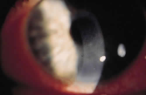
Many types of innate immunity occur in the eye. One example is the immune response to lipopolysaccharide or teichoic acids, found on microbe cell walls but not on mammalian cells. Recognition of lipopolysaccharide by CD4-positive lymphocytes leads to the release of tumor necrosis factor (TNF) and IL-12, activating neutrophils and natural killer cells leading to macrophage activation and inflammation.

SPECIFIC IMMUNE DEFENSES

Specific immune responses are classified into two types: humoral immunity, which is mediated by antibodies produced by B lymphocytes, and cell-mediated immunity (CMI), which is mediated by T lymphocytes.2

In humoral immunity, immunocompetent cells recognize an antigen and then must go to a central processing site, the lymphoid tissue, to produce antibodies. There are interrelated central processing sites throughout the body called mucous membrane-associated lymphoid tissues (e.g., in the lungs, gastrointestinal tract, and lacrimal gland). They lead to antibody distribution in all sites despite antigenic insult at only one site. Recently, Knop and Knop17 described conjunctiva-associated lymphoid tissue that contained all components, including follicular spots, to be necessary for a complete immune response. Before the demonstration of conjunctiva-associated lymphoid tissue, the predominant teaching was that in human eyes, after antigen sensitization in the mucosa-associated lymphoid tissue (Peyer's patches of the intestine), immunoglobulin A (IgA) committed precursor B cells and sensitized T cells to reach the general circulation and eventually reside in the lacrimal gland.18 Beneath the surface epithelium of the lacrimal gland, the B cells undergo further antigen stimulation and become plasma cells that secrete IgA and IgG.

Immunoglobulins are present at a higher concentration in tears than in serum.7 However, the concentration of IgA in tears is directly proportional to the tear flow rate. A diminished tear flow rate leads to increased IgA concentration while a person sleeps. This higher concentration of IgA can protect the ocular surface from residual organisms by neutralizing toxins and viruses and inhibiting the adherence of bacteria to mucosal surfaces, thus limiting an organism's ability to colonize the eye.19

Cell-mediated immunity also plays an important role locally and systemically in the eye's defense against microorganisms. When a T lymphocyte becomes sensitized to a bacterial antigen, it releases a soluble factor (lymphokine) that can invest the macrophage with the power to destroy ingested organisms. The sensitized T lymphocyte also releases factors that can aggregate macrophages at the site of insult and hinder their departure from the site. Chemotactic factors for neutrophils, basophils, and eosinophils are also released.2 The leukocyte infiltrate may confine the pathogen and prevent its entrance into the interior of the eye, but may also contribute to the necrotizing inflammation of the corneal stroma seen in gram-negative bacteria and herpes simplex virus (HSV).20

In CMI, several cytokines and interleukins play a role. IL-2 and its receptor have been found to be important mediators of the ocular inflammatory response, and a recent study by Nussenblatt and coworkers used IL-2 humanized anti-Tac antibody (ZenapaxÞRM [daclizumab], Hoffman La Roche, Nutley, NJ) in patients with chronic noninfectious uveitis.21 The mechanism of action of cyclosporin is also related to the inhibition of the production and release of IL-2. IL-2 is necessary for the induction of cytotoxic T lymphocytes in response to alloantigenic challenge and plays a major role in cellular and humoral immune response. Chemokines are produced by nearly all human cells. IL-8 has been shown to selectively induce neutrophil and eosinophil chemotaxis and degranulation in response to HSV and adenovirus.22

Back to Top
OFFENSIVE STRATEGIESOF THE ORGANISM
For ocular infection to occur, microorganisms must penetrate the host's barriers and be able to multiply without overly harming the host from whom it must derive shelter and nutrients. Exploitation of a host's intrinsic regulatory functions is an obvious way for microbes to accomplish this goal. Among an organism's offensive strategy are adherence, extracellular products to aid with invasion, and prevention of its own destruction by becoming unrecognizable or by blocking phagocytosis.

The first offensive strategy of bacteria is adherence to the host cell. Pili are fibrous organelles on the surface of gram-negative bacteria that aid in the adherence of the bacteria to the cells.23 They are required for colonization and are critical in the first steps in the development of disease.24 Pili are produced by plasmids (R factors), which can also contribute directly to virulence by converting the organism to phagocytosis-resistant forms. R factors are transferable between organisms and lead to one microorganism carrying combinations of different copies of R factors and contribute to antibiotic resistance.25

Staphylococcus aureus uses a different technique to adhere to the corneal surface. Certain strains of S. aureus produce proteins called adhesins, which have specific collagen binding sites. One study showed that adhesins aided in the pathogenesis of keratitis, especially with the use of contact lenses.26

Many bacteria contain extracellular products that help the organism to invade tissue. Gram-positive organisms invade tissue with the help of hyaluronidase, which depolymerizes hyaluronic acid.27 Group A streptococci produce fibrinolysins and streptokinase to digest fibrinogen and lyse fibrin clots. S. aureus produces alpha-toxin, lipase, protein A, exotoxins (extracellular bacterial toxins), leukocidins (bacterial toxins that destroy polymorphonuclear leukocytes), and coagulases (enzymes that accelerate the formation of blood clots), all of which help to invade the tissue. Other microbes produce exotoxins that are toxic to all cells of the reticuloendothelial system and help in tissue breakdown and invasion.28

Another technique that microbes use is to hide from the immune system and antibiotics. When antibiotics are present, some microorganisms, such as S. aureus, can also undergo transition to L forms, which lack the cell wall susceptible to antibiotics. Once the antibiotic has been withdrawn, the organism can reemerge as fully virulent.29 L-phase organisms bear strong resemblance to mycoplasma in their resistance to penicillin, lack of cell wall, and colony morphology.30 Structural lipids prevent destruction of certain organisms, including mycobac-teria, Listeria species, monocytogenes, and Francisella tularensis, from phagocytic digestion even once they are inside the phagolysosomes. The lipopolysaccharide endotoxins of gram-negative bacteria are a major constituent of their cell walls. Although innate immunity responds to lipopolysaccharide, alterations in the structure of lipopolysaccharide impair the ability of the host to respond to certain gram-negative bacteria.

Another method of preventing destruction is by blocking phagocytosis. S. aureus often produces fibrin-containing abscesses that act as an osmotic barrier to prevent the entrance of immunoglobulins into the focus of infection. Much of the surface of each S. aureus organism is covered with a unique substance called protein A, which helps to prevent phagocytosis of the bacterium.31 The antibody-binding fragment (Fab), which is normally the combining end of the antibody molecule, cannot combine with protein A; only the opposite end, the complement-binding fragment (Fc), combines with it. This reversed attachment blocks phagocytosis by macrophages and polymorphonuclear leukocytes. However, it also activates macrophages to release IL-1, increase phagocytosis, and increase TNF-alpha.31 Other microbes have other types of antiphagocytic factors as integral parts of their structure, for example, pneumococcal capsules and the M proteins of streptococcal cell walls that repel the host phagocytes by negative chemotaxis.32 The M proteins also bind complement regulatory and inhibitory factors leading to suppression of the complement pathway.33

Back to Top
NORMAL OCULAR FLORA
Under normal conditions, eyes interact with many microorganisms. The eyelid skin is colonized with nonpathogenic organisms similar to those in the upper respiratory tract, on the skin, and elsewhere. Under normal conditions, there is no growth of microorganisms from conjunctival cultures in 70% of people.34 The 30% of people who do have growth from conjunctival cultures generally are colonized with the same organisms that grow on the eyelids and eyelashes. These include gram-positive organisms, such as Staphylococcus species, Streptococcus species, Propionobacterium acnes, and Corynebacterium species, and gram-negative organisms, such as Haemophilus influenzae, Moraxella species, and Neisseria meningitides.7 Occasionally, Enterobacteriaceae species are found in elderly persons.35 The nonpathogenic organisms prevent the reintroduction of the pathogenic organisms.

GRAM-POSITIVE BACTERIA

Ocular bacterial infections can be caused by various organisms. Gram-positive cocci, including staphylococci and streptococci, account for the largest number of ocular infections.36 Gram-positive bacilli, including Bacillus cereus, Corynebacterum species, Listeria species, Clostridium species, and P. acnes, can also cause ocular infection. Because the cocci account for a much higher proportion of infections, they are discussed in greater detail.

Staphylococcus aureus is a gram-positive coccus that causes most cases of bacterial conjunctivitis and many of the culture-positive corneal ulcers.13 The traditional findings in S. aureus keratitis are a well-defined abscess with stromal infiltrates from the leukocytes attracted to fight infection. Clinical findings from staphylococcal blepharitis include poliosis, madarosis, and trichiasis, which reflects direct damage from the organisms to the glands of Zeis or the meibomian glands and the body's response (inflammatory and immunologic) to the microorganisms and their products (Fig. 2). In addition, ocular manifestations of S. aureus can be the result of type III and IV hypersensitivity reactions. The various presentations with this organism show the complex interaction of the immune system with different factors of the infecting organism.

Fig. 2. Ulcerated lid margin in a patient with staphylococcal blepharitis.

Staphylococcus aureus produces lipase to invade tissue. Decreasing the amount of lipase production with the use of tetracycline can aid in the treatment of blepharitis even at doses that do not decrease the growth of the bacteria.37

Staphylococci can cause immune-related disease with type III hypersensitivity reactions. Type III hypersensitivity is caused by an abnormal amount of antibody-antigen immune complexes deposited in the involved tissue. When antigens such as ribitol teichoic acid38 from the staphylococci bind to antibodies, neutrophils are attracted and can damage the tissues in the vicinity. This destruction manifests as white infiltrates at the limbus with excavation from damage to the cornea from the leukocyte products. The limbal distribution of these infiltrates results from the positioning of the lid margin and the presence of the appropriate amount of antigen and antibody in this area, caused by the lack of blood flow in the center of the cornea (Fig. 3). The infiltrates may regress without scarring if no necrosis has occurred, or it may progress to a breakdown of the overlying epithelium and finally to a peripheral ulcer. The lesions were once known as catarrhal ulcers, which is a misnomer because the destroyed cornea does not produce mucus, but the denuded surface may have increased adherence to mucus produced by goblet cells in the conjunctiva.

Fig. 3. Peripheral corneal ulceration in a case of S. aureusblepharitis.

Type IV hypersensitivity may also be seen with S. aureus in the form of phlyctenules. They represent CD4-positive delayed type hypersensitivity to a microbial antigen. Worldwide, most phlyctenules are caused by hypersensitivity to the tubercle bacillus,39 but in the United States, the lesion is more often associated with staphylococcal blepharoconjunctivitis. Other reported causes are Candida albicans, Coccidiodes immitis, chlamydia, and nemotodes. Phlyctenules are seen clinically as elevated limbal lesions that migrate toward the central cornea with a leash of vessels (Fig. 4). Late in the 10- to 14-day course, the nodular lesion ulcerates. Scarring is limited to the cornea and leaves a pathognomonic limbus-based triangular scar with the apex pointing toward the center of the cornea. Phlyctenules are more common in the first two decades of life.39 This can be explained by the high degree of cellular immunity in the early years of life.40 Another characteristic feature of severe cases is a wedge-shaped or fascicular pannus, found more often inferiorly than superiorly.

Fig. 4. Phlyctenules in a patient with chronic staphylococcal blepharitis.

Streptococci are responsible for a significantpercentage of ocular infections. Streptococcus pneumoniae can cause corneal ulceration through cytolysin, a protein that activates and releases degradative enzymes.41

Streptococcus viridans has been reported to cause infectious crystalline keratitis. This type of keratitis is often slow to respond to antibiotics because of the organism's production of a biofilm. A biofilm is an exopolysaccharide glycocalyx polymer that allows for protection from humoral and cellular immunity. It can also allow for protection from surfactants and antibiotics through dilution of the antibiotic and binding of antibiotics by exopolymers.42 The glycocalyx also blocks the interaction of the Fc receptor on host immune cells with antibody bound to bacterial surface antigens.43 A recent case report showed that disrupting the biofilm with an yterrium-aluminum-garnet photodisruptor can make the organisms available to the immune system for destruction.44

Other gram-positive bacilli, including B. cereus, Corynebacterium species, Listeria species, Clostridium species, and P. acnes, can cause keratitis or ocular infection. B. cereus is a ubiquitous organism and can be seen after a perforating injury. Corynebacterium species can cause pseudomembranous conjunctivitis. Listeria species can colonize persistent epithelial defects and lead to a ring ulcer formation. Clostridium species are distinct because of their formation of intrastromal gas and frothy bullous keratitis. P. acnes is a part of the normal flora and can cause indolent disease after surgery, trauma, or contact lens wear.45

GRAM-NEGATIVE BACTERIA

Gram-negative ocular infections are less common than gram-positive infections but can be more aggressive. One of the most devastating is Pseudomonas aeruginosa (Fig. 5); other gram-negative bacilli causing ocular disease include Acinetobacter species, a number of Enterobacteriaceae species, Neisseria species, Moraxella species, Haemophilus species, Mycobacterium species, and Nocardia species. Anaerobic bacilli, including Fusobacterium species, Bacteroides species, and Capnocytophagia species, can also cause ocular infection.

Fig. 5. Severe pseudomonal keratitis associated with contact lens wear.

Pseudomonas aeruginosa, a notorious cause of bacterial keratitis, can be derived from contaminated contact lens care material. The alterations in surface mucus with contact lens wear increases the susceptibility of the cornea to infection and allows P. aeuruginosa to adhere to the corneal surface.46 The aggressive nature of this organism in the cornea has been attributed in part to the production of an enzyme that destroys the proteoglycan that insulates and maintains the interfibrillar attachments of the corneal collagen.47

Meningococcus species, Gonococcus species, Haemophilus species, and other bacteria can produce membranous conjunctivitis as a result of the severity of the infectious process. A true membrane is a massive exudation of fibrin and proteinaceous fluid that permeates the conjunctival epithelium and superficial substantia propria and produces coagulative necrosis and subsequent cicatrization. The cornea is often affected secondarily.13

Capnocytophagia species are gram-negative rods normally found in human ocular flora. Although it is rare (0.3% in one study),48 it has an aggressive nature and poor outcome. Factors contributing to its virulence include the elicitation of an aggressive necrotizing inflammatory process and a protease that can degrade IgA and polyclonal IgG.48

Initially considered saprophytic for humans, Serratia marscens is a gram-negative rod that can cause ocular infection.49 It is known that the organism produces cornea-damaging proteases, but whether the ocular disease now apparently being caused by S. marscens is the result of altered host resistance, altered virulence of the microorganism, lack of past recognition, contaminated soft contact lens solution,50 or a combination of these and other factors is not known.

Back to Top
CHLAMYDIA
There are three species of chlamydia: Chlamydia trachomatis, Chlamydia psittaci, and Chlamydia pneumoniae. C. psittaci causes guinea pig inclusion conjunctivitis and has been studied extensively as a model for human eye disease. C. pneumoniae is the most recently described species. C. trachomatis is the most clinically significant cause of trachoma. Trachoma is highly endemic in some developing countries. It affects 300 to 400 million people and has blinded at least 6 million people.

The usual response of the conjunctival tissues to bacterial infections is papillary, but the chlamydial agents usually evoke a follicular response in the bulbar and palpebral conjunctiva. Rasmussen and colleagues51 showed that human endocervical cells produce IL-1 and IL-8 in the presence of chlamydia, indicating that CMI participates in the defense of the host against chlamydia agents. IL-8 is the most likely stimulus for recruitment of neutrophils to the local site. Although IgG and IgM have been detected in the sera of experimentally infected animals, it appears that any resistance to infection offered by antibodies is attributable to locally formed secretory IgA. It has been well shown that chlamydial infections will resolve spontaneously and that limited immunity to reinfection develops. During reinfection, fewer and fewer organisms are isolated on subsequent infections. To prevent the potentially blinding corneal scars, however, chlamydia requires prompt treatment.

Back to Top
SPIROCHETES
Syphilis is caused by the spirochete Treponema pallidum. Ocular findings can be seen in secondary syphilis in the form of iritis or anterior uveitis.52 Circulating immune complexes containing treponemal antigens and fibronectin combined with antibody and complement are present in this stage of infection and are thought to be responsible for the clinical findings.53 Syphilis may also present as interstitial keratitis in the congenital form (Fig. 6) or as a late finding in secondary syphilis with corneal scarring.54 There is a cell-mediated and humoral response to T. pallidum. The humoral response consists of nonspecific antibodies (reagins) that react to cardiolipin and specific antibodies that react to specific components on treponemes. These responses lead to an edematous cornea diffusely infiltrated with lymphocytes.49

Fig. 6. Interstitial keratitis.

Lyme disease, caused by the spirochete Borrelia burgdorferi, is a multisystem disorder heralded by a pathognomonic rash and neurologic and cardiac manifestation. There are three stages in Lyme disease: infection, dissemination, and late immunologic reactions. Each stage contains different ophthalmic findings. Stage I includes conjunctivitis and periorbital edema. Stage II has blepharospasm, iridocyclitis and uveitis, panophthalmitis, choroiditis, disc edema, and anterior ischemic optic neuropathy. Stage III includes stromal and interstitial keratitis, myositis, and cortical blindness.55 Ocular disease in stages I and II has been shown to include antigen-antibody and complement reactivation, delayed type hypersensitivity and vasculitis. Stage III is caused by a lasting autoimmune reaction secondary to antigenic mimicry.12

Back to Top
FUNGI
Fungi are ubiquitous organisms and have been isolated from up to 28% of healthy eyes.56 Various reports in the literature attribute fungi as the cause in 6% to 50% of cases of ulcerative keratitis (Fig. 7). More than 70 genera of filamentous fungi and yeasts have been identified in fungal keratitis. Nonpigmented fungi such as Fusarium species, Aspergillus species, Acremonium species, and Penicillium species cause most cases of fungal keratitis,57 seen after steroid use, trauma with vegetable matter, or insult by a deepithelialized cornea in chronic herpetic keratitis.

Fig. 7. Fungal keratitis with feathery stromal infiltrates.

The inflammatory reaction in fungal keratitis results from replicating and nonreplicating fungi, mycotoxins, proteolytic enzymes, and soluble fungal antigens.58 Filamentous fungi proliferate without the release of chemotactic substances that usually act to limit corneal damage.12 The immune response is mostly cell mediated, and high levels of IL-4, IL-10, and IgE have been found during the acute infection.10 Sequestration of fungal organisms under an intact epithelium allows the organisms to hide from topical antifungals.56

Back to Top
HERPESVIRUSES
There are seven distinct types of herpesviruses. All are morphologically similar, but they have substantial differences in their structural glycoproteins and polypeptides. The herpesvirus family includes two types of HSV, cytomegalovirus, varicella zoster virus, Epstein-Barr virus, and human herpesviruses 6 and 7.14

Herpes simplex is a visually devastating virus and is the leading cause of infectious blindness in the United States.59 Humans are the only natural host of HSV. Herpetic infection of the eye is the result of direct invasion of ocular tissue by the virus (Fig. 8). In chronic herpes simplex keratitis, the herpesvirus is latent and the herpetic antigenic determinants are incorporated into the cell walls of infected trigeminal ganglion cells. These cells become chronic antigenic sources and may stimulate immune reactions without active infection. When an appropriate trigger mechanism (e.g., fever, stress, and ultraviolet light) activates HSV in a ganglion,12 the virus is shed into the tear film, and an infectious recurrence follows. The trigger mechanism may cause a transient depression in the immune system leading to the virus having the opportunity to reactivate.

Fig. 8. Dendrite with adherent mucus in a patient with primary infection with herpes simplex virus.

Once the herpes virus is active, anterior segment damage ensues. HSV may possess collagenolytic activity, which would account for some of its virulence, but the corneal damage is thought to be mainly caused by the immune response rather than direct virus-induced damage. Neutrophils are thought to play a crucial role in tissue damage and may perpetuate corneal inflammation by exposing neoantigens subject to T-cell responses and by the release of IL-12.12 In mice, it has been found that IL-1α (a proinflammatory cytokine) and TNF-α (synergistic with IL-1) are upregulated in recurrent herpes simplex keratitis.59 Among the functions of IL-1 are to upregulate adhesion molecules, enhance neovascularization, and serve as a cofactor in lymphocyte activation.60 IL-10, however, may play a role in controlling keratitis induced by herpes simplex.61 Mice infected with HSV-1 had decreased local viral growth and decreased corneal opacification after receiving topical IL-10.62

The clinical manifestations of herpes simplex depend on the immune status of the host. Iritis is unusual except in immunosuppressed hosts. Disciform keratitis may be a manifestation of CMI, with the herpetic antigen sensitizing T lymphocytes, which may then cause the edematous, inflammatory condition (Fig. 9). Because virus may be present in the uveal tract, the inflammatory response of the uveal tissue may be the result of the virus directly or the immunologic defenses.

Fig. 9. Disciform keratitis with a leash of vessels surrounded by clear cornea.

Immune globulin has been used to treat HSV, but it has resulted in an increased incidence of disciform keratitis, probably a manifestation of antigen-antibody reaction. The use of immunopotentiators like topical interferon for the treatment of ocular viral infections has been considered for more than 30 years. At this point there has been no proven effective use of immunopotentiators for HSV.

Herpes zoster virus can manifest clinically in various ways. The incidence of infection with herpes zoster virus increases by 2% to 4% per year of life.63 This is probably related to the decreasing cellular immune response. Chronic lymphocytic infiltration can cause tissue damage by inflammation and vasculitis-induced ischemia. Disciform keratitis is the result of delayed type hypersensitivity.64

The Epstein-Barr virus can cause ocular disease by infection of B cells. The various clinical manifestations associated with Epstein-Barr virus include Parinaud oculoglandular syndrome, conjunctivalinflammation, Sjogren syndrome, keratitis, and uveitis.13

NONHERPETIC VIRUSES

More than 40 species of viruses can cause disease in the eye. Fortunately, most viral infections (i.e., adenoviral, enteroviral, and picornaviral) are in the form of self-limited conjunctivitis. Immunotherapy (vaccination) is a successful way of preventing potentially devastating viral infections, such as measles, mumps, and rubella.

Adenoviruses are a common cause of benign conjunctivitis. There are more than 77 distinct species of adenovirus.65 Each adenovirus particle contains at least three morphologic subunits (the major capsid protein, the fiber, and the penton) that contribute to its immunologic reactivity. The major capsid protein contains an antigen that cross-reacts with a comparable antigen in all adenoviruses. The fiber, which allows the virus to attach to host cells, cannot induce synthesis of neutralizing antigens. The complex intact penton is a minor antigen that serves biologically as a cytopathic factor and an endonuclease.65 Epidemic keratoconjunctivitis (caused most commonly by adenoviruses 7, 8, 19, and 37) is the only adenoviral syndrome associated with corneal inflammation. The corneal infiltrates have been shown to be caused by polymorphonuclear neutrophils in early stages and later to be caused by lymphocytes (Fig. 10).22

Fig. 10. Subepithelial infiltrates caused by viral conjunctivitis.

Because the use of antivirals with corticosteroids for any form of ocular virus disease has the potential for many complications resulting from prevention of the healing process and immunosuppression of the host defense systems (e.g., increased viral multiplication and secondary infection), the therapeutic role of corticosteroids remains controversial.66 The role of corticosteroids in any ocular infection should be questioned except in the small number of cases in which antimicrobial agents can effectively destroy all the infecting microorganisms while the corticosteroids protect visually important structures from damage that may occur during the inflammatory response of the reparative process. Inflammation is not disease; rather, it is one of the body's major defenses against disease and an indispensable defense against the opportunistic pathogen. In general, corticosteroids do not have a role in acute infection but may aid in chronic sterile postviral inflammation.66

Back to Top
PARASITES
There are more than 30 types of parasites that have been shown to cause ocular infection.8 In many cases of parasitic infection, the inflammatory reaction causes a greater problem than the infecting organism itself.

Acanthamoeba species are ubiquitous, free-living, freshwater protozoa that reside in soil, tap water, fresh and brackish seawater, dust, swimming pools, and contact lens cases.67 Contact lens wearers who use homemade saline or who swim with contact lenses account for 85% of patients with Acanthamoeba keratitis (Fig. 11).68 Causes not related to contact lens wear include trauma and topical steroid use. The pain and chemosis are often disproportionate to the normal inflammatory process.8 There may be a radial neurokeratitis, or an opaque circular infiltrate from type III hypersensitivity with immune complex deposition may develop late in the course of the infection. This circle is known as the Wessely immune ring and is also seen in HSV infection (Fig. 12). It has been shown pathologically to be a result of immune complex deposition with local fixation of complement in the cornea and release of pharmacologic agents that cause chemotaxis of polymorphonuclear leukocytes that surround the complexes.20 The Wessely immune ring can also be seen in HSV and fungal keratitis.

Fig. 11. Disciform keratitis from Acanthamoeba infection.

Fig. 12. Wessely immune ring caused by the “zone of optimal proportions” of antibody and antigen (courtesy of Jeffrey Lanier, M.D., Houston, TX).

Histologically, when amoebae are demonstrated, it is usually with mild inflammation.34 There may be no demonstrable amoebae in the zone of greatest inflammation, leading to the concept that the living trophozoites do not incite host reactions because of a protective mechanism of the parasite that assumes a nearly indestructible nonmotile cyst when stressed,1 but that the dying cysts release antigens that were previously hidden.68 This makes the eradication of Acanthamoeba species from the ocular tissue difficult.67

Oncocerca volvulus, the filarial nematode that causes river blindness and leads to various ocular lesions, including stromal keratitis and chorioretinitis, may cause an allergic reaction to the parasite antigen. Eosinophils and fibrinoid material, shown to be immune complex deposition, have been found coating these worms.8 In the mouse model of the disease, infiltration of T lymphocytes, predominantly CD3- and CD4-positive during the development of the lesions.69 There is an upregulation of IL-4 and IL-5, but IL-2 and TNF-τ were not increased.69 IL-4 has several proinflammatory activities, including increased expression of vascular adhesion molecule-1 on endothelial cells. IL-5 is responsible for eosinophil development, migration, and activation. It is thought that the major basic protein released from the eosinophil granule is partly responsible for the disease leading to blindness in onchocerciasis.

Ocular cysticercosis, caused by tapeworm species Taenia solium and Taenia saginata, causes only mechanical compression with mild fibrosis in the early cases. When the worm dies, there is a severe inflammatory reaction to the toxic products of the parasites.8

Back to Top
OPPORTUNISTIC INFECTIONS
Resistance to all pathogens is lowered in patients who are old, malnourished, debilitated, ill with Hodgkin disease, atopic dermatitis, diabetes mellitus, AIDS, and other diseases and in patients who are artificially immunosuppressed (e.g., by immunosuppressive therapy after transplant surgery). This can lead to the emergence of ocular infections caused by organisms previously regarded as saprophytic. Examples are the central corneal ulcers now being caused by α-streptococci and by mycotic and parasitic organisms. However, in one study that evaluated microbial flora in immune deficient patients, the flora did not differ greatly from normal flora. Patients with B-cell deficiencies, in particular, may have a high incidence of infectious disease affecting the eye.70 CMI is a key protective mechanism in fungal infections. One example of this is in endogenous endophthalmitis resulting from C. albicans. Under normal circumstances, C. albicans is part of the normal skin, reproductive tract, and gastrointestinal microbial flora,12 and the immune system keeps it at a tolerable level. This is partly the result of the lack of outstanding virulence factors that C. albicans has. In the absence of CMI or in patients with weak granulocyte function (e.g., patients taking steroids and patients with AIDS or diabetes), C. albicans can enter the bloodstream and cause systemic disease, including endophthalmitis.12

Atopic dermatitis is a chronic pruritic inflammatory skin disease characterized by local infiltration of monocytes and T cells, mast cell degranulation, and a combination of immediate and cellular immune response.12 Interestingly, the inflamed skin of patients with atopic dermatitis has increased attachment sites for S. aureus, and reduced skin inflammation is associated with reduced colonization.71 Bonifazi and coworkers found that patients with atopic dermatitis were more susceptible to infection caused by a T-lymphocyte deficiency, specifically T-suppressor cells.72 The T-cell deficiency in patients with atopic dermatitis leads to impaired CMI and a higher likelihood of developing corneal stromal lesions with HSV, and those with intact T-cell function usually develop only epithelial lesions (Fig. 13).73Molluscum contagiosum can also take advantage of defective T-cell function and cause conjunctival and corneal lesions.74

Fig. 13. Corneal ulcer in a patient with atopic dermatitis, chronic herpetic keratitis, and bacterial keratitis.

In AIDS there are more than a dozen types of infections that become more prevalent when a patient is deficient in CD4-positive cells. Patients with AIDS have a more severe course with organisms in each category previously discussed: bacteria, spirochetes (tertiary syphilis), viruses, and parasites. Varicella zoster virus is reactivated in patients with varying levels of immune compromise. It has been shown to occur at a much greater rate in patients with HIV than in age-matched control subjects. As noted earlier, Molluscum contagiosum also runs rampant when there is a T-cell deficiency. It can even present on the ocular surface, as reported by Ingraham and coworkers.74 Microsporidia are obligate intracellular protozoa that cause keratitis, primarily in patients with AIDS. Presentation includes epiphora, photophobia, conjunctival injection, and punctate epithelial erosions. Diagnosis is made with conjunctival scrapings that show aggregates of gram-positive, intracellular, oval organisms. Because of the persistent susceptibility of the patient to this organism, topical fumagilin may need to be used every hour indefinitely.75

Back to Top
AREAS FOR FUTURE FOCUS
As new surgical procedures continue to develop, there will be new scenarios in which the immune system can react to infection in the eye. For example, diffuse lamellar keratitis, which, of course, was first described after the advent of laser-assisted in situ keratomileusis has been attributed to be caused by various factors, including the immune response to toxins from bacteria in a biofilm containing reservoir and blepharitis (Fig. 14). As new surgical procedures, pharmacologic products, and situations arise, there will always be new and interesting circumstances for the eye to interact with the immune system.

Fig. 14. Diffuse lamellar keratitis after laser-assisted in situ keratomileusis in a patient with blepharitis.

Back to Top
ACKNOWLEDGMENTS
The authors thank Dr. Sami Uwaydat and Dr. Nelson C. Klaus III for their suggestions during the preparation of this manuscript. Bruce and Arlene Tonjes were also invaluable for their editorial and style contributions. The authors also thank Dr. G. Smolin, who wrote the previous edition of this chapter; we acknowledge his contribution to the framework of this manuscript.
Back to Top
REFERENCES

1. Silverstein AM: Ocular immunology: on the birth of a new discipline. Cell Immunol 136:504–518, 1991.

2. Abbas AK, Lichtman AH, Pober JS. Cellular and molecular immunology, 4th ed. Philadelphia: WB Saunders, 2000:viii, 553.

3. Ohta K, Yamagami S, Taylor A et al: IL-6 antagonizes TGF-beta and abolishes immune privilege in eyes with endotoxin-induced uveitis. Invest Ophthalmol Vis Sci 41:2591–2599, 2000.

4. Zierhut M, Bieber T, Brocker EB et al: Immunology of the skin and the eye. Immunol Today 17:448–450, 1996.

5. Streilin JW, Yamada J, Dana MR et al: Anterior chamber-associated immune deviation, ocular immune privilege, and orthotopic corneal allografts. Transplant Proc 31:1472–1475, 1999.

6. Friedlaender MH: Allergy and immunology of the eye, 2nd ed. New York: Raven Press, 1993:ix, 337.

7. Smolin G: The defence mechanism of the outer eye. Trans Ophthalmol Soc UK 104:363–366, 1985.

8. Kean BH, Sun T, Ellsworth RM: Color atlas/text of ophthalmic parasitology. New York: Igaku-Shoin, 1991:xiv, 233.

9. Arnold RR, Russell JE, Champion WJ et al: Bactericidal activity of human lactoferrin: differentiation from the stasis of iron deprivation. Infect Immunol 35:792–799, 1982.

10. O'Day DM: New antifungal drugs in ophthalmology. Int Ophthalmol Clin 36:45–51, 1996.

11. Gachon AM, Lacazette E: Tear lipocalin and the eye's front line of defence. Br J Ophthalmol 82:453–455, 1998.

12. Cunningham MW, Fujinami RJ: Effects of microbes on the immune system. Philadelphia, Lippincott Williams & Wilkins, 2000:xix, 662.

13. Kaufman HE, Barron BA, McDonald MB: The cornea, 2nd ed. Boston, Butterworth-Heinemann, 1998:xxi, 1109.

14. Ryan KJ, Sherris JC: Sherris medical microbiology: an introduction to infectious diseases, 3rd ed. Norwalk, CT, Appleton & Lange, 1994:xiii, 890.

15. Zasloff M: Antibiotic peptides as mediators of innate immunity. Curr Opin Immunol 4:3–7, 1992.

16. Haynes RJ, McElveen JE, Dua HS e al: Expression of human beta-defensins in intraocular tissues. Invest Ophthalmol Vis Sci, 41:3026–3031, 2000.

17. Knop N, Knop E: Conjunctiva-associated lymphoid tissue in the human eye. Invest Ophthalmol Vis Sci 41:1270–1279, 2000.

18. Friedman MG: Antibodies in human tears during and after infection. Surv Ophthalmol 35:151–157, 1990.

19. Zierhut M, Elson CO, Forrester JV et al: Mucosal immunology and the eye. Immunol Today 19:148–150, 1998.

20. Meyers-Elliott RH, Pettit TH, Maxwell WA: Viral antigens in the immune ring of herpes simplex stromal keratitis. Arch Ophthalmol 98:897–904, 1980.

21. Nussenblatt RB, Schiffman R, Fortin E et al: Strategies for the treatment of intraocular inflammatory disease. Transplant Proc 30:4124–4125, 1998.

22. Chodosh J, Astley RA, Butler M et al: Adenovirus keratitis: a role for interleukin-8. Invest Ophthalmol Vis Sci 41:783–789, 2000.

23. Soto GE, Hultgren SJ: Bacterial adhesins: common themes and variations in architecture and assembly. J Bacteriol 181:1059–1071, 1999.

24. Sauer FG, Mulvey MA, Schilling JD et al: Bacterial pili: molecular mechanisms of pathogenesis. Curr Opin Microbiol 3:65–72, 2000.

25. Amyes SG: The success of plasmid-encoded resistance genes in clinical bacteria. An examination of plasmid-mediated ampicillin and trimethoprim resistance genes and their resistance mechanisms. J Med Microbiol 28:73–83, 1989.

26. Rhem MN, Lech EM, Patti JM et al: The collagen-binding adhesin is a virulence factor in Staphylococcus aureus keratitis. Infect Immunol 68:3776–3779, 2000.

27. Hynes WL, Walton SL: Hyaluronidases of gram-positive bacteria. FEMS Microbiol Lett 183:201–207, 2000.

28. Rhen M, Eriksson S, Pettersson S: Bacterial adaptation to host innate immunity responses. Curr Opin Microbiol 3:60–64, 2000.

29. Paton AM: L-forms: evolution or revolution? J Appl Bacteriol 63:365–371, 1987.

30. Butler HM, Blakey JL: A review of bacteria in L-phase and their possible clinical sigificance. Med J Aust 2:463–467, 1975.

31. Ghosh AK, Sinha P, Das T et al: S. aureus superantigen protein A expands CD4(+ )/CD8(+ )/CD19(+ )/CD34(+ ) cells in mice: a potential immunorestorer. Biochem Biophys Res Commun 256:142–146, 1999.

32. Rautemaa R, Meri S: Complement-resistance mechanisms of bacteria. Microbes Infect 1:785–794, 1999.

33. Johnsson E, Thern A, Dahlback B et al: A highly variable region in members of the streptococcal M protein family binds the human complement regulator C4BP. J Immunol 157:3021–3029, 1996.

34. McClellan KA: Mucosal defense of the outer eye. Surv Ophthalmol 42:233–246, 1997.

35. Seal DV: Bacterial classification and diagnosis. Trans Ophthalmol Soc UK 105:32–36, 1986.

36. Dajcs JJ, Hume EB, Moreau JM et al: Lysostaphin treatment of methicillin-resistant Staphylococcus aureus keratitis in the rabbit. Invest Ophthalmol Vis Sci 41:1432–1437, 2000.

37. Dougherty JM, McCulley JP, Silvany RE et al: The role of tetracycline in chronic blepharitis. Inhibition of lipase production in staphylococci. Invest Ophthalmol Vis Sci 32:2970–2975, 1991.

38. Mondino BJ, Laheji AK, Adamu SA: Ocular immunity to Staphylococcus aureus. Invest Ophthalmol Vis Sci 28:560–564, 1987.

39. Rohatgi J, Dhaliwal U: Phlyctenular eye disease: a reappraisal. Jpn J Ophthalmol 44:146–150, 2000.

40. Ostler HB, Lanier JD: Phylctenular keratoconjunctivitis with special reference to the staphylococcal type. Trans Pac Coast Otoophthalmol Soc Annu Meet 55:237–252, 1974.

41. Johnson MK, Allen JH: The role of cytolysin in pneumococcal ocular infection. Am J Ophthalmol 80:518–521, 1975.

42. Elder MJ, Stapleton F, Evans E et al: Biofilm-related infections in ophthalmology. Eye 9:102–109, 1995.

43. Grasso RJ, Ganguly R, Breen JF: Inhibition of yeast phagocytosis in macrophage cultures treated with slime polysaccharide purified from Pseudomonas aeruginosa. J Leukoc Biol 36:771–774, 1984.

44. Daneshvar H, MacInns B, Hodge WG: Nd:YAG laser corneal disruption as adjuvant treatment for infectious crystalline keratopathy. Am J Ophthalmol 129:800–801, 2000.

45. Yanoff M, Duker JS, eds. Ophthalmology. London, Mosby, 1999:1.

46. Lyczak JB, Cannon CL, Pier GB: Establishment of Pseudo-monas aeruginosa infection: lessons from a versatile oppor-tunist. Microbes Infect 2:1051–1060, 2000.

47. Fleiszig SM, Zaidi TS, Ramphal R et al: Modulation of Pseudomonas aeruginosa adherence to the corneal surface by mucus. Infect Immun 62:1799–1804, 1994.

48. Alexandrakis G, Palma LA, Miller D et al: Capnocytophagia keratitis. Ophthalmology 107:1503–1506, 2000.

49. Parment PA, Ronnerstam RA: Soft contact lens keratitis associated with Serratia marcescens. Acta Ophthalmol (Copenh) 1981:560–565, 1981.

50. Parment PA: The role of Serratia marcescens in soft contact lens associated ocular infections. A review. Acta Ophthalmol Scand 75:67–71, 1997.

51. Rasmussen SJ, Eckmann L, Quayle AJ et al: Secretion of proinflammatory cytokines by epithelial cells in response to Chlamydia infection suggests a central role for epithelial cells in chlamydial pathogenesis. J Clin Invest 99:77–87, 1997.

52. Albert DM, Jakobiec FA: Principles and practice of ophthalmology, 2nd ed, vol 6. Philadelphia, WB Saunders, 2000:5583.

53. Baughn RE, McNeely MC, Jorizzo JL et al: Characterization of the antigenic determinants and host components in immune complexes from patients with secondary syphilis. J Immunol 136:1406–1414, 1986.

54. Knox CM, Holsclaw DS: Interstitial keratitis. Int Ophthalmol Clin 38:183–195, 1998.

55. Winterkorn JM: Lyme disease: neurologic and ophthalmic manifestations. Surv Ophthalmol 35:191–204, 1990.

56. Mabon M: Fungal keratitis. Int Ophthalmol Clin 38:115–123, 1998.

57. Garg P, Gopinathan U, Choudhary K et al: Keratomycosis: clinical and microbiologic experience with dematiaceous fungi. Ophthalmology 107:574–580, 2000.

58. Zhu WS, Wojdya K: Extracellular proteases of Aspergillus flavus. Fungal keratitis, proteases, and pathogenesis. Diagn Microbiol Infect Dis 13:491–497, 1990.

59. Keadle TL, Usui N, Laycock KA et al: IL-1 and TNF-alpha are important factors in the pathogenesis of murine recurrent herpetic stromal keratitis. Invest Ophthalmol Vis Sci 41:96–102, 2000.

60. Staats HF, Lausch RN: Cytokine expression in vivo during murine herpetic stromal keratitis. Effect of protective antibody therapy. J Immunol 151:277–283, 1993.

61. Tumpey TM, Elner VM, Chen SH et al: Interleukin-10 treatment can suppress stromal keratitis induced by herpes simplex virus type 1. J Immunol 153:2258–2265, 1993.

62. Minagawa H, Sakai Y, Li Y et al: Suppression of infectious virus spread and corneal opacification by the combined use of recombinant interferon beta and interleukin-10 following corneal infection with herpes simplex virus-1 in mice. Antiviral Res 36:99–105, 1997.

63. Ragozzino MW, Melton LJ, Kurland LT et al: Population-based study of herpes zoster and its sequelae. Medicine (Baltimore) 61:310–316, 1982.

64. McGill J: The enigma of herpes stromal disease. Br J Ophthalmol 71:118–125, 1987.

65. Ginsberg HS: Adenoviruses. Am J Clin Pathol 57:771–776, 1972.

66. Baratz KH, Hattenhauer MG: Indiscriminate use of corticosteroid-containing eyedrops. Mayo Clin Proc 74:362–366, 1999.

67. Pineda R II, Dohlman CH: The role of steroids in the management of Acanthamoeba keratitis, fungal keratitis, and epidemic keratoconjunctivitis. Int Ophthalmol Clin 34:19–31, 1994.

68. Illingworth CD, Cook SD: Acanthamoeba keratitis. Surv Ophthalmol 42:493–508, 1998.

69. Chakravarti B, Lagoo-Deenadayalan S, Parker JS et al: In vivo molecular analysis of cytokines in a murine model of ocular onchocerciasis. I. Up-regulation of IL-4 and IL-5 mRNAs and not IL-2 and IFN gamma mRNAs in the cornea due to experimental interstitial keratitis. Immunol Lett 54:59–64, 1996.

70. Friedlaener MH, Masi RJ, Osumoto M et al: Ocular microbial flora in immunodeficient patients. Arch Ophthalmol 98:1211–1213, 1980.

71. Stalder JF, Fleury M, Sourisse M et al: Local steroid therapy and bacterial skin flora in atopic dermatitis. Br J Ophthalmol 131:536–540, 1994.

72. Bonifazi E, Garofalo L, Pisani V et al: Role of some infectious agents in atopic dermatitis. Acta Derm Venereol Suppl 114:98–100, 1985.

73. David TJ, Longson M: Herpes simplex infections in atopic eczema. Arch Dis Child 60:338–343, 1985.

74. Ingraham HJ, Schoenleber DB: Epibulbar molluscum contagiosum. Am J Ophthalmol 125:394–396, 1998.

75. Silverstein BE: Parasitic corneal infections. Int Ophthalmol Clin 38:179–182, 1998.

Back to Top