Chapter 33
Immunology of Rheumatoid Diseases That Affect the Eye
MITCHELL H. FRIEDLAENDER
Main Menu   Table Of Contents

Search

ANKYLOSING SPONDYLITIS
REITER'S SYNDROME
RHEUMATOID ARTHRITIS
JUVENILE RHEUMATOID ARTHRITIS
SYSTEMIC LUPUS ERYTHEMATOSUS
SJO¯GREN'S SYNDROME
POLYARTERITIS NODOSA
WEGENER'S GRANULOMATOSIS
GIANT CELL ARTERITIS
POLYMYOSITIS-DERMATOMYOSITIS
PROGRESSIVE SYSTEMIC SCLEROSIS (SCLERODERMA)
REFERENCES

Ocular disease is a characteristic feature of many rheumatologic disorders. Autoimmune mechanisms are believed to play a significant role in a number of these conditions, and a variety of immunologic abnormalities connected with rheumatoid disease already have been identified. Because the eye is only one of many organs affected, the immunologic abnormalities known to characterize the disease in other organs should throw light on the pathogenesis of the ocular lesions as well.
Back to Top
ANKYLOSING SPONDYLITIS
Ankylosing spondylitis is a chronic, progressive, inflammatory arthritis that affects principally the sacroiliac joints and spine. About 25% of cases are associated with an acute, recurrent iridocyclitis, often bilateral, and accompanied by synechiae and glaucoma. Males are affected with ankylosing spondylitis more often than females, accounting for 80% to 90% of all patients. The disease occurs most frequently in the second and third decades of life. Some 90% of patients have the histocompatibility antigen HLA-B27. This represents the highest correlation observed so far between an HLA type and a disease entity.

IMMUNOPATHOLOGY

The cause of ankylosing spondylitis is unknown. For many years, it was considered a variant of rheumatoid arthritis but is now known to differ in several important respects. There is no strong evidence of an autoimmune mechanism, and rheumatoid factor is negative except in approximately 5% of cases. It is well known, however, that genetic factors play a significant role. The disease is 40 times more common among first-degree relatives than in the general population, and the concordance rate in affected identical twins is 70%.1 Family pedigrees suggest that the gene for ankylosing spondylitis is inherited as a Mendelian dominant trait with 70% penetrance in males and 10% in females.2 It also is known that the frequency of ankylosing spondylitis is high among certain American Indian groups such as the Haida, Bella Coola, and Pima and relatively rare among blacks.3,4

Histocompatibility (HLA) antigens are cell surface determinants present on most human cell membranes. Their specificity in humans is governed by a group of genes located on the sixth chromosome. Each gene determines a cell surface antigen so that an individual will have four cell surface antigens that are determined genetically. The frequency of an HLA type can be determined by testing a patient's lymphocytes with a number of different antisera.

Although HLA typing has been used principally to match donors and recipients in organ transplantation, the association of HLA types with specific disease entities is now receiving considerable attention. In 1973, Schlosstein and his colleagues5 in Los Angeles and Brewerton and his colleagues6 in England reported a strikingly high incidence of HLA-B27 in patients with spondylitis. They found the antigen (which has a frequency of 4% to 9% in control populations) in 95% of patients with ankylosing spondylitis, and comparably high percentages have been reported by other investigators.

Ankylosing spondylitis is common in families that propagate the HLA-B27 antigen. It has been estimated that if the risk that an antigen-negative female will have spondylitis is one, then the risk in a B27-positive female is 276 times greater. For a B27-positive male, the risk is 1937 times greater.

The significance of this high frequency of HLA-B27 in patients affected with ankylosing spondylitis is uncertain. The antigen also is found in 90% of patients with a spondylitis that occurs after infection with Yersinia enterocolitica. This is a gram-negative bacterium that causes a transient inflammatory bowel disorder that is sometimes followed by polyarthritis and sacroiliitis. In patients who recover uneventfully from the infection, the incidence of HLA-B27 is the same as for the general population, but of those in whom arthritis develops, B27 is found in approximately 90%. This suggests that spondylitis can follow an infectious disease in a genetically susceptible individual. Whether this is because of a defective immune response or a hypersensitivity mechanism is not known. An infectious disease process also may be the basis of Reiter's syndrome and even of ankylosing spondylitis.

Back to Top
REITER'S SYNDROME
Reiter's syndrome consists of a triad of lesions—acute conjunctivitis, nonspecific urethritis, and arthritis—with punctate keratitis, corneal stromal opacities, and iritis, sometimes also in evidence. The disease usually occurs in sexually active males and often follows dysenteric infections with Shigella, Yersinia, or Salmonella.

IMMUNOPATHOLOGY

As with ankylosing spondylitis, most patients with Reiter's syndrome (90%) have the histocompatibility antigen HLA-B27. The reason for this association is not known. The first possibility is that the B27 antigen may act as a favorable receptor site for certain microbial pathogens, a number of which, especially Chlamydia, Mycoplasma, and Shigella, have been irregularly associated with Reiter's syndrome. A second possibility is that infectious agents that cause Reiter's syndrome and other rheumatoid diseases share common antigens with the host's own connective tissues. This could result in the host's inability to recognize and combat the invading microbe, or alternatively it could result in an immume response directed against the host's tissues as well as against the infectious agent. A third possibility, and the most widely accepted, is that HLA-B27 is linked in the genetic material to an immune-response gene and that this gene is responsible for the clinical syndrome. The immune response may be directed appropriately against the infecting agent or inappropriately against the patient's own tissues.

There are only a few immunologic abnormalities in patients with Reiter's syndrome. Cellular immunity is intact, although lymphocyte transformation has been shown with chlamydial and prostatic antigens.7 In some patients with Reiter's syndrome, cell-mediated immunity to autologous immunoglobulin G (IgG) also has been found.8

In one of many attempts that have been made to implicate a microorganism in Reiter's syndrome, Schachter and associates9 propagated a chlamydial agent that they had isolated from synovial fluid, synovial membranes, urethras, and conjunctival of patients with the syndrome. When they recovered the organism from the synovial membrane of an affected patient and inoculated it into the anterior chambers of rabbits, ocular disease (consisting of papillary conjunctivitis, corneal edema, corneal opacities, corneal neovascularization, and iritis) developed.10 Several cases of Reiter's syndrome also have been reported in association with Shigella dysentery and other enteric infections, and it may be that a number of infectious agents can produce the syndrome in genetically susceptible individuals.11

Back to Top
RHEUMATOID ARTHRITIS
Rheumatoid arthritis is a chronic, recurrent, systemic inflammation that affects mainly the peripheral joints. It occurs in 2% or 3% of the population, is three times more frequent in females than in males, and usually appears between the ages of 30 and 50 years. Constitutional symptoms may include malaise, fever, and weight loss. The peripheral joints on both sides of the body are affected first, and the arthritis usually progresses centripetally. Scleritis is a frequent and important complication, and the cornea and uveal tract may be affected secondarily. There is no consistent pattern of inheritance in rheumatoid arthritis and no known prevalence of any HLA antigen.

IMMUNOPATHOLOGY

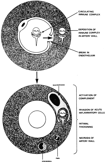
The antigenic stimulus that initiates joint inflammation in rheumatoid arthritis still is a mystery. Certain microorganisms have been implicated, but their causal roles still are uncertain. Viruses, particularly the slow viruses, may play a role, although no virus particles have been certainly identified. The results of attempts to isolate Mycoplasma and rubella virus from the joints of patients with rheumatoid arthritis have been inconclusive.12 During the prodromal stage of hepatitis, hepatitis B virus can form immune complexes and produce a syndrome resembling serum sickness with polyarthralgia and vasculitis. This virus seems to be the first virus known to produce a chronic rheumatic disorder in humans, and, like many other infectious agents, it can give rise to rheumatoid factor in serum. The clinical syndrome, however, bears no resemblance to either rheumatoid arthritis or systemic lupus erythematosus (SLE).

Other possible infectious causes of rheumatoid arthritis have been investigated. Rheumatoid synovial cells show a diminished sensitivity to infection with either Newcastle disease virus or rubella virus. Mycoplasma antibodies have been isolated from patients with rheumatoid arthritis, especially from those with longstanding disease, and recently a slow-growing infectious agent with some of the properties of Mycoplasma has been isolated from the synovial fluid of such patients.13 In other studies, however, investigators have failed to find any evidence of previous Mycoplasma infection in patients with rheumatoid arthritis.14

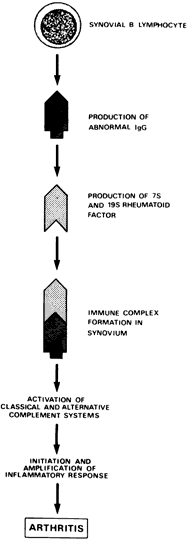
A variety of immunologic abnormalities have been found in patients with rheumatoid arthritis, and there now is considerable evidence that the disease is caused by an autoimmune process. Antibodies against IgG are formed in the patient's blood and synovial fluid. Immune complexes that are formed and deposited in the joints and other tissues activate the complement system through the classic and alternate pathways.15 Activation of the complement system results in a number of inflammatory phenomena, including chemotaxis of leukocytes, histamine release, and cell lysis. Enzymes released by the synovial leukocytes produce inflammatory changes in the joints and the destruction of normal structures. The inflammatory response is amplified by the various Immoral amplification systems.

The Immoral immune system appears to be highly active and important in the pathogenesis of rheumatoid arthritis (Fig. 1). The number of synovial B lymphocytes, which are precursors of antibody-producing plasma cells, often is abnormally high. More than 50% of the synovial plasma cells produce IgG rheumatoid factor, an antibody directed against other IgG molecules.10 Immune complexes may be found within plasma cells of the synovial membrane, a finding unparalleled in any other immunopathologic disorder. Although a greater-than-normal number of peripheral B cells usually are found, the number is hard to estimate because of the antilymphocyte antibodies present.16 When these antibodies are removed, the number of peripheral B cells may in fact be reduced.

Fig. 1. Hypothetic immunopathogenesis in rheumatoid arthritis. (Fye KH, Moutsopoulos HM, Talal N: Rheumatoid diseases. In Fudenberg HH, Stites DP, Caldwell JL et al [eds]: Basic and Clinical Immunology, p 366. Los Altos: Lange Medical Publications, 1976.)

The following antibodies also have been identified in the sera of patients with rheumatoid arthritis: (1) antibodies to double-stranded DNA; (2) antibodies to human native and denatured collagen; and (3) antinuclear antibodies (ANAs).17,18 Recently, it has been shown that IgG molecules in the sera of patients with rheumatoid arthritis have a conformational anomaly in the hinge region.19 This altered IgG may be recognized as abnormal by B-lymphocyte receptors, leading to an Immoral autoimmune response directed against IgG.

The following defects in cellular immunity also have been associated with rheumatoid arthritis:

  1. When tested with multiple skin-test antigens, 20% of affected patients are anergic.20
  2. Although some investigators have reported abnormally low levels of peripheral blood T lymphocytes during active disease, others have found the levels slightly elevated. In synovial fluid from actively inflamed joints, they have been consistently high.21,22
  3. Heat-aggregated IgG and, to a lesser extent, native IgG inhibit the migration of rheumatoid leukocytes.

This T-cell response to IgG antigens and the fact that soluble mediators of lymphocytes contribute to the inflammatory changes that take place in the rheumatoid joint strongly suggest that there is a cell-mediated immune component in rheumatoid arthritis.

A third population of lymphocytes, lacking conventional B- and T-cell markers, may be important in the pathogenesis of rheumatoid disease. This population, known as “null cells,” may include the so-called killer lymphocytes (K cells) that are cytotoxic to IgG-coated target cells.22 Null cells may be responsible for the formation of “rheumatoid rosettes,” which are formed by the interaction of lymphocytes and IgG-coated indicator erythrocytes. In addition, peripheral blood leukocytes collected from patients with rheumatoid arthritis may be cytotoxic for synovial cells.23

Antigen preparations of uvea-retina, synovial membrane, and articular cartilage inhibit the migration of leukocytes obtained from patients with rheumatoid arthritis.24 In ankylosing spondylitis, inhibition is induced only by synovial membrane antigens. Lymphocytes from blood and synovial fluid of patients with rheumatoid arthritis also show a markedly diminished blastogenic response to phytohemagglutinin and pokeweed mitogen.25

The reason for depressed cellular immunity in rheumatoid arthritis is unknown. It may result from a preoccupation of the host's immune mechanism with cell-mediated immune reactions related to the pathogenesis of the disease, or it may be related to a systemic viral infection. Alternatively, depressed cellular immunity may be caused by immune complex formation or by rheumatoid arthritis therapy. The HLA-Dw4 allele occurs with high frequency and DQw7 influences severity.26

Back to Top
JUVENILE RHEUMATOID ARTHRITIS
Juvenile rheumatoid arthritis (JRA) is a major chronic, progressive, crippling disease of childhood. It has a peak incidence between the ages of 2 and 4 years and a lesser peak between the ages of 10 and 12 years. It occurs twice as often in females as in males and has an average duration of approximately 6½ years. A major complication of JRA is chronic iridocyclitis, and cataract formation and band keratopathy also may occur. The ocular disease is one of the leading causes of uveitis in childhood and may be associated with significant visual disability. JRA is conveniently divided into three forms: (1) the acute, toxic form (Still's disease); (2) a polyarticular form; and (3) a pauciarticular form. Patients with this third form have the highest incidence of iridocyclitis.

IMMUNOPATHOLOGY

Although rheumatoid factor is prevalent in adult rheumatoid arthritis, it is not commonly found in patients with JRA, with only 10% to 20% testing positively compared with 50% to 85% of those with the adult disease. Conversely, ANAs are found in 20% to 40% of children with JRA (most with pauciarticular and polyarticular JRA but a few with Still's disease) and in 88% of JRA patients who have chronic iridocyclitis.27,28 The ANAs are predominantly of the IgG immunoglobulin class and the titers are 1:50 or higher in most patients. The consistent homogeneity of the nuclear fluorescence pattern is typical of reactivity against deoxyribonucleoprotein.29 Antibodies to DNA and RNA usually are not demonstrable, but antibodies against double-stranded RNA recently have been reported in children with JRA and iridocyclitis.30 A positive ANA test result usually precedes the onset of iridocyclitis and may be useful in identifying patients with JRA who are likely to have chronic iridocyclitis develop. The reason for a positive ANA test result in this disease is not known, but in general both infectious processes and host immune defects are associated with the formation of ANA and other autoantibodies.

In patients with JRA and iridocyclitis, smooth-muscle antibodies, usually of the immunoglobulin M (IgM) class, are found in 15% to 23%, which is the same frequency with which they are found in control groups. The HLA antigens of patients with JRA have been examined, and some investigators have reported a high incidence of HLA-B27.31,32 Others have not found this to be the case, however, and suggest that the earlier studies included patients with ankylosing spondylitis, which, in its early stages, can mimic JRA.33,34

The clinical picture of Still's disease suggests the possibility of a disseminated infection. Evidence indicating an infectious etiology is scant, however. A rise in antibody titer to Coxsackie B3 and A9 viruses and the isolation of adenovirus 7 have both been reported.29 The titer of rubella virus antibody has been found to be elevated, and rubella virus antigen has been isolated from synovial fluid. Perhaps a viral infection, combined with defective immunity, allows an infectious agent to persist in eye and joint tissues, which leads to the development of JRA.

Back to Top
SYSTEMIC LUPUS ERYTHEMATOSUS
Systemic lupus erythematosus is a multisystem collagen-vascular disease. The etiology is unknown, but hereditary, immunologic, and microbiologic factors seem to be concerned. SLE is characterized by a tendency to form autoantibodies, especially to constituents of the cell nucleus. The main organ systems affected are the joints, skin (including the skin of the eyelids), kidneys, lungs, and heart. The eye and central nervous system also may be affected. Cotton-wool spots and retinal vasculitis may occur and sometimes scleritis, corneal opacities, and optic neuritis. SLE is seen most commonly in women of the child-bearing age and more commonly in nonwhites than in whites.

IMMUNOPATHOLOGY

Recent investigations have shown that genetic susceptibility, possible viral infection, and abnormal immune responses interact in the pathogenesis of SLE. Antibody formation to double-stranded DNA occurs frequently and is associated with disease activity. Depressed T-cell activity, enhanced B-cell activity, and a low level of serum complement also are prominent features of the disease.

Much of our current understanding of SLE is derived from animal studies in which an animal model of the disease in New Zealand black (NZB) mice was used. NZB mice have a deficiency of suppressor T cells and a related low level of a circulating thymic hormone known as thymosin. They are immunologically impaired and susceptible to various infections, including those caused by oncogenic viruses. Both NZB mice and hybrid NZBNZW (New Zealand white) mice have been studied extensively. NZB mice, hybrid mice, and humans with SLE have ANAs in their blood and deposits of both immune complexes and components of the complement system in their kidneys.

These immune complexes contain DNA and antibodies against DNA, but the source of the DNA is not known. It could be released from damaged cells (possibly as a result of viral infection) or it could itself be of viral origin. Viruses have been implicated in the development of both human SLE and the mouse disease. Type C virus has been identified in NZB mice and can be transmitted genetically from parent to progeny in the egg and sperm. Copies of the viral genome can thus be incorporated into the cellular genome. It has been suggested that a genetic defect in NZB mice permits expression of the viral genome and that this, in turn, results in autoimmune disease. The pathologic changes might be caused by the development of antibodies against viral components, with the antibodies forming immune complexes with the viral antigen and being deposited in the kidneys and other organs. A second possibility is that viral antigens on cell surfaces provoke an immunologic attack on these infected cells, which are not recognized by the host as “self.” Antigens related to those of type C virus also have been identified on the lymphocytes and in the kidneys of some patients with SLE. Fifty to 60% of patients have antibodies to cardiolipin.35 This antibody may be associated with occlusive retinal vasculitis.

Currently, the most popular conception of the pathogenesis of SLE is as follows: The infectious agent is a type C RNA virus, which is known to be transmitted genetically in animals. Because of a genetic predisposition of the host, the virus avoids the immune surveillance system and homes in on, replicates, or is recognized in the thymus. The thymic alteration may eventually destroy T-cell function, either directly or by way of an autoimmune mechanism. Further viral replication and dissemination are permitted because of a defect in cellular immunity, and this leads to an escalated humoral immune response. Immune complexes are formed, circulated, and deposited in various organs, producing inflammatory reactions and activation of the complement system. Inflammation and clinical disease then develop.

Studies of lymphocytes from patients with SLE indicate that when the disease is active, the T cells are markedly reduced, the null cells are increased, and the B cells usually remain at their normal level. T-cell lymphopenia may be caused by the increased number of null cells. (The null cells, which mature on exposure to thymus, may simply be immature T cells. If this is the case, the population of T cells would increase as the null cells mature.)

Monocyte-macrophage function is depressed in early SLE, and since these cells participate in the processing of antigen and in lymphokine activity, this defect could result in depressed cellular immunity.36 A lupus-like syndrome has been associated with deficiency of the second component of complement (C2), and it may be that an inherited deficiency of this component could predispose an individual to lupus or to faulty handling of viral infections.37

Histocompatibility antigens have been investigated in both SLE and discoid lupus erythematosus (DLE), a cutaneous variant of SLE. Female patients with SLE show a significant association with HLA-B8, and patients with DLE have a greater-than-normal association with both HLA-B7 and HLA-B8.38

Back to Top
SJO¯GREN'S SYNDROME
Sjögren's syndrome is a chronic inflammatory disease of unknown origin that primarily affects the lacrimal and salivary glands. Its clinical hallmark is dryness of the oral mucosa, conjunctive, and other mucous membranes. Half of the patients with Sjögren's syndrome have rheumatoid arthritis, and a smaller group have other connective tissue diseases. A presumptive diagnosis of Sjögren's syndrome can be made if two of the following three features are present: (1) recurrent or chronic idiopathic swelling of the salivary glands; (2) unexplained xerostomia; and (3) connective tissue disease. A definitive diagnosis can be made if there is objective evidence of keratoconjunctivitis sicca or a typical histopathologic change in the lacrimal or salivary glands.

IMMUNOPATHOLOGY

Sjögren's syndrome is characterized by the development of lymphocytic infiltrates and by destruction of the salivary and lacrimal glands and other tissues. Both T and B lymphocytes have been detected in the salivary gland infiltrates.39 Examination of the peripheral blood usually shows a modest increase in the peripheral B lymphocytes, and the T lymphocytes are reduced in approximately one-third of patients. The lymphocytes infiltrating the salivary glands synthesize IgM and IgG (locally), rheumatoid factor, and other autoantibodies.

Hypergammaglobulinemia, usually of the polyclonal type, is the most frequent immunopathologic finding in Sjögren's syndrome. Rheumatoid factor is found in at least 75% of patients, and the titers are especially high in those with the sicca complex (keratoconjunctivitis sicca and xerostomia alone). ANAs occur in approximately 90% of patients, and the staining pattern usually is homogeneous or speckled.40 Antidouble-stranded DNA antibodies and lupus erythematosus (LE) cells are found in a small percentage of patients, and autoantibodies against salivary-duct antigens may be detected in approximately 50% of patients.

Whereas humoral antibody formation in Sjögren's syndrome is exuberant, cellular immunity appears to be somewhat depressed. Patients with the disease do not have delayed hypersensitivity to contact allergens develop, and peripheral blood lymphocytes do not respond normally to mitogens.41 Cellular hypersensitivity to salivary gland extract has been demonstrated by the use of peripheral blood leukocytes from affected patients. Perhaps T-lymphocyte sensitization to salivary gland antigens (with the production of lymphokines) occurs and is responsible for pathologic changes. As in other autoimmune disorders, there probably is a defect in T-suppressor cells in Sjögren's syndrome. Such a deficiency could permit the uncontrolled proliferation of B cells and the development of overactive immune responses such as autoantibody formation. Antibodies to Epstein-Barr virus have been detected in the blood and saliva of patients with Sjögren's syndrome.42

Patients with Sjögren's syndrome are predisposed to such lymphoproliferative disorders as lymphoma, leukemia, and Waldenstrom's macroglobulinemia.43 An intermediate stage of lymphoproliferation, known as “pseudolymphoma,” also may occur. Whether Sjögren's syndrome is the result of a slow or latent viral disease or a disorder of immunologic regulation remains to be determined. Androgens and prolactin may play a role in regulating the lacrimal gland.44

Back to Top
POLYARTERITIS NODOSA
Polyarteritis nodosa (once known as periarteritis nodosa) is a connective tissue disorder and one of a number of arteritis-vasculitis syndromes; its hallmark is widespread inflammation of medium and small-sized arteries. Nearly any organ can be affected, and there are ocular manifestations—scleritis, sclerokeratitis, uveitis, retinal vasculitis, and optic neuritis—in 10% to 20% of patients. A number of factors suggest that the disease is immunologic in nature and probably mediated by immune complex deposition within vessels. Other clinical entities that can be considered arteritis-vasculitis syndromes are hypersensitivity angiitis, temporal arteritis, vasculitis associated with rheumatic fever, and allergic granulomatous angiitis. The pathogenesis of these syndromes (which were classified by Zeek45 and then more fully by Hawkins46 [Table 1]) is thought to be similar to that of polyarteritis nodosa.


TABLE 1. The Arteritis-Vasculitis Syndromes

  Polyarteritis Nodosa
  Hypersensitivity Angiitis

  Drug-induced
  Serum sickness arteritis
  Anaphylactoid purpura (Henoch-Sch<auo>nlein syndrome)


  Vasculitis Associated With Connective Tissue Diseases

  Rheumatoid arthritis
  Systemic lupus erythematosus
  Progressive systemic sclerosis
  Polymyositis, dermatomyositis
  Sj<auo>gren's syndrome


  Vasculitis Associated With Rheumatic Fever
  Vasculitis With a Granulomatous Component

  Allergic granulomatous angiitis (Churg and Strauss)
  Wegener's granulomatosis
  Lethal midline granuloma
  Limited Wegener's granulomatosis
  Lymphomatoid granulomatosis
  Giant cell or temporal arteritis
  Aortic arch syndrome (Tayayasu's disease)


  Other Vasculitides and Disorders Associated with Vasculitis

  Mixed cryoglobulinemia
  Drug abuse (methamphetamines)
  Hepatitis-associated antigenemia with vasculitis (Australia antigenemia)
  Nonsuppurative inflammatory bowel disease
  Postcoarctation resection
  Pulmonary hypertension
  Systemic hypertension
  Reticuloendothelial malignancies
  Goodpasture's syndrome
  Syphilis
  Erythema nodosum
  Nodular vasculitis
  Weber-Christian disease
  Cogan's syndrome


(Adapted from Zeek PM: Am J Clin Pathol 22:777, 1952.)


IMMUNOPATHOLOGY

Although most cases of polyarteritis nodosa appear to be related to hypersensitivity, the antigen (or antigens) has not been identified. However, humoral factors play a role. This is indicated by the following:

  1. Repeated intravenous injections of heterologous protein given to rabbits produce an arteritis similar to polyarteritis nodosa.
  2. Bovine gamma globulin administration not only induces a polyarteritis but can lead to immune complex deposition in the kidneys.
  3. The injection of immune complexes intravenously in rats is followed by vasculitis.

In human cases, immune complexes have been deposited in vessel walls during active disease. The work of Gocke and associates47 has shown that circulating immune complexes composed of Australia antigen and immunoglobulin are present in the sera of patients in whom polyarteritis nodosa has been established by biopsy. Some of these patients also show deposition of Australia antigen, IgM, and complement in blood vessel walls (Fig. 2). Although this intriguing finding suggests a relationship between polyarteritis nodosa and exposure to a microorganism, most cases do not have circulating immune complexes containing Australia antigen.

Fig. 2. Hypothetic immunopathogenesis in polyarteritis nodosa associated with Australia antigen. (Fye KH, Moutsopoulos HM, Talal N: Rheumatoid diseases. In Fudenberg HH, Stites DP, Caldwell JL et al [eds]: Basic and Clinical Immunology, p 380. Los Altos: Lange Medical Publications, 1976.)

Renal biopsy specimens from patients with the disease have shown deposition of such immune reactants as immunoglobulins, complement, and fibrin. Such deposition has not been found consistently, however, and may in fact be nonspecific.

Microbial antigens, drugs, and autoantigens may play a role in widespread vascular inflammation. Streptococcal antigens and numerous drugs—sulfonamides, penicillin, diphenylhydantoin, arsenical, thiouracil, iodides, and thiazides—have been associated with polyarteritis, hypersensitivity angiitis, and Henoch-Schonlein purpura. There also is a high association between methamphetamine abuse and polyarteritis nodosa. The fact that many such drug users eventually have hepatitis develop strengthens the evidence that there is an association between polyarteritis nodosa and Australia antigen.

Histologically, the typical lesion of polyarteritis nodosa is an infiltration of neutrophils and eosinophils around medium and small arteries. The necrotizing, inflammatory process is located most commonly at blood-vessel bifurcation sites. Round-cell infiltration occurs in the chronic phase of inflammation and is followed ultimately by fibrinoid necrosis.

Back to Top
WEGENER'S GRANULOMATOSIS
Wegener's granulomatosis usually is described as a triad of lesions: (1) necrotizing granulomas of the respiratory tract; (2) disseminated vasculitis of the small and medium-sized arteries and veins; and (3) glomerulonephritis. It affects mainly young and middle-aged individuals, and ocular lesions—orbital and nasolacrimal granulomas, peripheral corneal degeneration, scleritis, and retinal vasculitis—frequently occur. A limited form of the disease has been described in which the pulmonary lesions are typical, but lesions elsewhere are limited or absent. Midline granuloma is sometimes classified as a manifestation of Wegener's granulomatosis, but the validity of such a classification has been seriously questioned.

IMMUNOPATHOLOGY

Wegener's granulomatosis is a necrotizing vasculitis with the features of an autoimmune disorder. Circulating autoantibodies to smooth muscle, circulating immune complexes, and reduced levels of serum complement have all been found. Complement and immunoglobulins have been detected in the vascular lesions of the skin and kidneys.48 IgG and C3 were found in the glomeruli of one patient, suggesting an immune complex deposition, and IgG, C3, and fibrin were found in the glomerular vessels of another patient.49,50 Elevated levels of serum IgA, IgE, and C3 also have been reported.51 Subepithelial basement-membrane deposition of IgG and complement has been found in the glomerular tufts in a lumpy-bumpy pattern characteristic of immune complex disease.

The level of delayed skin hypersensitivity to various common skin-test antigens is abnormally low in Wegener's granulomatosis. Reduced lymphocyte mitogenic responses also have been documented, but some of the studies were performed on patients treated with immunosuppressive agents.

Back to Top
GIANT CELL ARTERITIS
Giant cell arteritis is a term used to describe a group of vasculitides in which epithelioid or giant cells infiltrate the walls of vessels. The principal diseases are temporal arteritis, polymyalgia rheumatica, and aortic-arch syndrome or Takayasu's disease, with temporal arteritis the most important to the ophthalmologist. Because of the ischemic optic neuritis and the central retinal-artery occlusion that sometimes accompany the disease, temporal arteritis ranks as one of the prime medical emergencies in ophthalmology. Rapid recognition and prompt treatment are essential for the preservation of vision in affected patients.

IMMUNOPATHOLOGY

The hallmark of temporal arteritis is granulomatous inflammation that affects the temporal arteries selectively.52 The presence of epithelioid or giant cells is characteristic. Destruction of the internal elastic lamina is common but not diagnostic. The best way to confirm the diagnosis is with a temporal artery biopsy. But although it has been stated that a pattern of inflammation characteristic of temporal arteritis can always be found in any such biopsy specimen, according to other observers, “skip areas” make it possible for an entire biopsy specimen to lack the tell-tale evidence.52,53

Immunofluorescent studies have shown deposition of IgG, IgA, IgM, and complement in the cytoplasm of cells and in the elastic tissue within vessel walls.54 ANA directed against nuclei of cells within vessel walls may be seen in patients who have serum ANA. A nonspecific elevation of the levels of alpha-2 globulin and fibrinogen in plasma also has been noted. Because these proteins reduce the electronegative charge on circulating erythrocytes and promote rouleaux formation, their elevation may account for the high erythrocyte sedimentation rate in patients with giant cell arteritis.55 Immunoglobulin abnormalities usually are not observed, but elevated levels of IgM have been noted in a number of patients.55 Lymphocytes from patients with polymyalgia rheumatica undergo blast transformation in vitro in the presence of either arterial antigens or homogenates of whole muscle contaminated with vascular antigens.54,56

There has been some recent speculation that viral antigens may participate in giant cell arteritis and polymyalgia rheumatica. Cytoplasmic inclusion bodies recently have been found in temporal artery biopsy specimens.57 The meningoencephalitis caused by varicella virus has been reported as having histologic features similar to those of giant cell arteritis, and others have found the vessels of the central nervous system selectively affected by varicella virus.57,58 Segmental artery involvement also has been reported shortly after an attack of herpes zoster ophthalmicus.59 Perhaps viruses or other infectious agents can alter vascular antigens and produce an autoallergic inflammation.

Takayasu's disease is associated with an elevation of serum IgG, IgA, and IgM.60 Perhaps there also is a fundamental immunologic abnormality in this disease.

Back to Top
POLYMYOSITIS-DERMATOMYOSITIS
Polymyositis and dermatomyositis encompass a spectrum of related disorders characterized by inflammation and degeneration of varying amounts of skeletal muscle. They are multisystem diseases with the features of rheumatic disorders and may occur in association with malignancy. Ocular manifestations rarely are reported but do occur. The most common are a heliotrope rash affecting the eyelids, iritis, conjunctivitis, episcleritis, cotton-wool spots, retinal hemorrhages, and extraocular muscle palsies. Five categories of polymyositis-dermatomyositis have been recognized: (1) idiopathic polymyositis; (2) idiopathic dermatomyositis; (3) polymyositis-dermatomyositis associated with malignancy; (4) childhood polymyositis-dermatomyositis; and (5) polymyositis-dermatomyositis associated with other rheumatoid diseases.

IMMUNOPATHOLOGY

The etiology of these disorders currently is unknown. Features of both humoral and cellular immunity have been recognized in affected patients, and there has been speculation that a virus may play an etiologic role.

Circulating antibodies to muscle can be found after any muscle injury. Local deposits of IgG, IgM, and complement have been found within the vessel walls of affected skin and muscle in polymyositis.61 These deposits, detectable mainly in children with diffuse vasculitis, suggest the possibility of an immune complex disorder. Polyclonal hypergammaglobulinemia often is seen in patients with polymyositis-dermatomyositis, and both rheumatoid factor and ANA are demonstrable in 20% of cases. Because patients with agammaglobulinemia can have polymyositis develop, immunoglobulins are clearly not a requirement.62 Dermatomyositis has been reported in association with C2 deficiency, indicating further that the classic pathway of complement activation is not always required.63 Deficiencies of the complement system have been reported in association with a number of rheumatic abnormalities, suggesting that patients with rheumatic disease are abnormally susceptible to immunologic injury.

There is evidence that cellular immunity participates in the development of polymyositis-dermatomyositis. Lymphocytes from patients with active myositis induce cytotoxic changes in autologous or chick muscle cells in tissue culture.64 Muscle cell injury is thought to be caused by lymphotoxin, a lymphokine produced by T cells. The lymphocytes from affected patients sometimes respond to their own muscle antigens as though they were foreign (Fig. 3). This could be because of an altered membrane antigen on the muscle cells, induced perhaps by a virus. Alternatively, there may be a primary defect in antigen recognition by lymphocytes or a cross-reactivity between muscle antigen and an unidentified foreign antigen. Presumably, the cellular response would occur at the site of the instigating antigen, and lymphotoxin could diffuse through the tissues and injure the remote muscle fibers.

Fig. 3. Defective recognition of muscle antigen in polymyositis. (Fye KH, Moutsopoulos HM, Talal N: Rheumatoid diseases. In Fudenberg HH, Stites DP, Caldwell JL et al [eds]: Basic and Clinical Immunology, p 377. Los Altos: Lange Medical Publications, 1976.)

Electron microscopy has identified virus-like particles of various structural types in the myocytes of patients with polymyositis. Although the etiology of these disorders remains speculative, a combination of viral and immunologic abnormalities is suggested by current available evidence. Histologic findings show an infiltration of muscle cells with a focal or diffuse lymphocytic infiltrate. Necrosis, degeneration, and regeneration of muscle cells are typical.

Back to Top
PROGRESSIVE SYSTEMIC SCLEROSIS (SCLERODERMA)
Progressive systemic sclerosis is a connective tissue disease affecting various organ systems, especially the skin, lungs, and gastrointestinal tract. For many years, it was considered primarily a dermatologic condition, but its systemic nature is now well recognized. The main pathologic feature is a greater-than-normal collagen deposition in the affected organs. The rare ocular findings cover a spectrum of abnormalities that include brawny edema of the eyelids, keratoconjunctivitis sicca, cotton-wool spots, retinal hemorrhage, disc edema, and uveitis.

IMMUNOPATHOLOGY

The etiology of progressive systemic sclerosis is unknown, but the following three pathogenic mechanisms may possibly contribute to the development of the disease: (1) an immunologic defect, (2) an autonomic nervous system defect, and (3) a primary connective tissue defect.

Several immunologic abnormalities have been detected in affected patients. Most patients have a hypergammaglobulinemia of the polyclonal type, and rheumatoid factor is found in approximately 35% of patients. Test results for lupus erythematosus cells occasionally are positive, and serologic test results for syphilis are falsely positive. ANAs, usually of the coarse, speckled, nuclear type, are present in at least 60% of patients but do not correlate with the clinical severity of the disease.65

Antinucleolar antibodies and antibodies to ribonucleoprotein also are found, and one investigating team recently found a new ANA marker system by using a concentrated tissue extract as antigen.66 This new antibody seems to be specific for the disease, since it is not found in other connective tissue disorders.

A mixed cryoglobulin containing IgG and IgM and usually associated with antigammaglobulin activity in serum has been found in 50% of patients with progressive systemic sclerosis.62 Although this suggests the possibility of circulating immune complexes, their role (if they exist) and the etiology of the disease remain uncertain. There still is relatively little evidence that the associated vascular abnormalities are based on a humoral immunologic defect.

There also is some evidence that cellular immune mechanisms participate in progressive systemic sclerosis. Lymphocytes from patients with the disease can destroy embryonic fibroblasts in tissue culture. Cellular infiltration of affected tissue is slight, however, except in the synovium.

The importance of a component of the autonomic nervous system in progressive systemic sclerosis has been emphasized.67 The high incidence of both Raynaud's phenomenon and esophageal abnormalities, their reversal by reserpine and methacholine, and their impaired responses to gastrin I and edrophonium all suggest an autonomic abnormality. To be effective, the latter two drugs (gastrin I and edrophonium) require an intact autonomic nervous system.

A primary defect in collagen has been suggested as contributing to progressive systemic sclerosis. Collagen biosynthesis is elevated in tissue culture, and there are abnormalities in the conversion of proline to hydroxyproline.68,69

By microscopic examination, sclerosis and fibrosis are seen in various tissues, and blood vessels may show intimal fibrosis, endothelial proliferation, and hyperplasia of the media. Although many facts are known about progressive systemic sclerosis, a unified, workable hypothesis that would account for its various clinical manifestations has not yet been put together.

Back to Top
REFERENCES

1. Jullcunen H: Rheumatoid spondylitis: Clinical and laboratory studies of 149 cases compared with 182 cases of rheumatoid arthritis. Acta Rheum Scan 4(Suppl):4, 1962

2. Emery AEH, Lawrence JS: Genetics of ankylosing spondylitis. J Med Genet 4:239, 1967

3. Gofton JP, Bennett PH, Smythe HA et al: Sacroileitis and ankylosing spondylitis in North American Indians. Ann Rheum Dis 31:474, 1972

4. Gofton JP, Lawrence JS, Bennett PH et al: Sacroileitis in eight populations. Ann Rheum Dis 25:528, 1966

5. Schlosstein L, Terasaki PI, Bluestone R et al: High association of an HLA antigen, W27, with ankylosing spondylitis. N Engl J Med 288:704, 1973

6. Brewerton DA, Caffrey M, Hart ED et al: Ankylosing spondylitis and HLA27. Lancet 1:904, 1973

7. Amor B, Kahan A, Lecoq F et al: Le test de transformation lymphoblastique par les antigenes bedsoniens (TTL Bedsonien). Rev Rheum Mal Osteoartic 39:671, 1972

8. Weisbart RH, Bluestone R, Goldberg LS: Cellular immunity to autologous IgG in rheumatoid arthritis and rheumatoidlike disorders. Clin Exp Immunol 20:409, 1975

9. Schachter J: Isolation of Bedsoniae from human arthritis and abortion tissues. Am J Ophthalmol 63:1082, 1967

10. Ostler HB, Schachter J, Dawson CR: Ocular infection of rabbits with a Bedsonia isolated from a patient with Reiter's syndrome. Invest Ophthalmol 9:256, 1970

11. Good AK, Schultz JS: Reiter's syndrome following Shigella flexneri 2a. Arthritis Rheum 20:100, 1977

12. Editorial. Lancet 2:79, 1976

13. Jansson E, Makisara P, Tuuri S: Mycoplasma antibodies in rheumatoid arthritis. Scand J Rheumatol 4:165, 1975

14. Cole BC, Taylor MB, Ward JR: Studies on the infectious etiology of human rheumatoid arthritis: II. Search for humoral and cellbound antibodies against mycoplasmal antigens. Arthritis Rheum 18:435, 1975

15. Zvaifler NJ: The immunopathology of joint inflammation in rheumatoid arthritis. Adv Immunol 16:265, 1973

16. Winfield JB, Winchester RJ, Wernet P et al: Antibodies to lymphocytes and the estimation of B and T cells in the connective tissue disorders. In Beers RF, Bassett EG (eds): The Role of Immunological Factors in Infectious, Allergic, and Autoimmune Processes, p 137. New York: Raven Press, 1976

17. Bell C, Talal N, Schur PH: Antibodies to DNA in patients with rheumatoid arthritis and juvenile rheumatoid arthritis. Arthritis Rheum 18:535, 1975

18. Andriopoulos NA, Mestecky J, Miller EJ et al: Antibodies to human native and denatured collagens in synovial fluids of patients with rheumatoid arthritis. Clin Immunol Immunopathol 6:209, 1976

19. Johnson PM, Watkins J, Holborow EJ: Antiglobulin production to altered IgG in rheumatoid arthritis. Lancet 1:611, 1975

20. Andrianakos AA, Sharp JT, Person DA et al: Cell-mediated immunity in rheumatoid arthritis. Ann Rheum Dis 36:13, 1977

21. Williams RC Jr, Debord JR, Mellbye OJ et al: Studies of B and T lymphocytes in patients with connective tissue diseases. J Clin Invest 58:690, 1976

22. Freland SS, Natvig JB, Wisloff F et al: Lymphocyte reactions in rheumatoid arthritis. In Beers RF Jr, Bassett EG (eds): The Role of Immunologic Factors in Infectious, Allergic, and Autoimmune Processes, p 289. New York: Raven Press, 1976

23. Person DA, Sharp JT, Lidsky MD: Cytotoxicity of leukocytes and lymphocytes from patients with rheumatoid arthritis for synovial cells. J Clin Invest 58:690, 1976

24. Thonar EJMA, Sweet MBE: Cellular hypersensitivity in rheumatoid arthritis, ankylosing spondylitis, and anterior nongranulomatous uveitis. Arthritis Rheum 19:539, 1976

25. Hepburn B, McDuffie FC, Ritts RE Jr: Impaired blastogenic response of lymphocytes from synovial fluid and peripheral blood of patients with rheumatoid arthritis. J Rheumatol 3:118, 1975

26. Harper S, Foster CS: Ocular manifestations of rheumatoid arthritis. Int Ophthalmol Clin 33:1, 1998

27. Schaller JG, Wedgwood RJ: Is juvenile rheumatoid arthritis a single disease? A review article. Pediatrics 50:940, 1972

28. Schaller JG, Johnson GD, Holborow EJ et al: The association of antinuclear antibodies with the chronic iridocyclitis of juvenile rheumatoid arthritis (Still's disease). Arthritis Rheum 17:409, 1974

29. Friou GJ: Antinuclear antibodies: Diagnostic significance and methods. Arthritis Rheum 10:151, 1967

30. Epstein WV, Tan EM, Easterbrook M: Serum antibody to double-stranded RNA and DNA in patients with idiopathic and secondary uveitis. N Engl J Med 285:1502, 1971

31. Buc M, Nyulassy S, Stefanovic J et al: HLA system and juvenile rheumatoid arthritis. Tissue Antigens 4:395, 1974

32. Rachelefsky G, Teraski PI, Katz R et al: Increased prevalence of W27 in juvenile rheumatoid arthritis. N Engl J Med 290:892, 1974

33. Mitsui H, Juji T, Sonozaki H et al: Distribution of HLAB27 in patients with juvenile rheumatoid arthritis. Ann Rheum Dis 36:86, 1977

34. Ohno S, Char D, Kimura SJ et al: HLA antigens and antinuclear antibody titers in juvenile chronic iridocyclitis. Br J Ophthalmol 61:59, 1977

35. Rosenbaum JT: Systemic lupus erythematosus. In FT Fraunfelder, FH Roy (eds): Current Ocular Therapy, p 173. Philadelphia: Saunders, 1999

36. Landry M: Phagocyte function and cell-mediated immunity in systemic lupus erythematosus. Arch Dermatol 113:147, 1977

37. Douglass MC, Lamberg SI, Lorincz AI et al: Lupus erythematosus-like syndrome with a familial deficiency of C2. Arch Dermatol 112:671, 1976

38. Millard LG, Rowell NR, Rajah SM: Histocompatibility antigens in discoid and systemic lupus erythematosus. Br J Dermatol 96:139, 1977

39. Recent clinical and experimental developments in Sjögren's syndrome. Medical staff conference, University of California San Francisco. West J Med 122:50, 1975

40. Block KJ, Buchanan WW, Wohl MJ et al: Sjögren's syndrome: A clinical, pathological, and serological study of sixty-two cases. Medicine 44:187, 1965

41. Shearn MA: Sjogrens syndrome. Semin Arthritis Rheum 2:165, 1972

42. Mircheff AK, Gierow JP, Wood RL: Autoimmunity of the lacrimal gland. Int Ophthalmol Clin 34:1, 1994

43. Cummings NA, Schall GL, Asofsky R et al: Sjögren's syndrome: Newer aspects of research, diagnosis, and therapy. Ann Intern Med 75:937, 1971

44. Warren DW: Hormonal influences on the lacrimal gland. Int Ophthalmol Clin 34:19, 1994

45. Zeek PM: Periarteritis nodosa: A critical review. Am J Clin Pathol 22:777, 1952

46. Hawkins D: Diffuse connective tissue diseases: I. Systemic lupus erythematosus and related disorders, pp 256–294. In Freedman SO, Gold P (eds): Clinical Immunology, 2nd ed. Hagerstown: Harper & Row, 1976

47. Gocke DJ, Hsu K, Morgan C et al: Vasculitis in association with Australia antigen. J Exp Med 134:330S, 1971

48. Hu CH, O'Laughlin S, Winkelman RK: Cutaneous manifestation of Wegener's granulomatosis. Arch Dermatol 113:175, 1977

49. Fauci AS, Wolff SM: Wegener's granulomatosis: Studies in eighteen patients and review of the literature. Medicine 52:535, 1973

50. Roback SA, Herdman RC, Hoyer J et al: Wegener's granulomatosis in a child: Observations on pathogenesis and treatment. Am J Dis Child 112:587, 1966

51. Conn DL, Gleich GJ, DeRemee RA et al: Raised serum immunoglobulin E in Wegener's granulomatosis. Ann Rheum Dis 35:377, 1976

52. Albert DM, Ruchman MC, Keltner JL: Skip areas in temporal arteritis. Arch Ophthalmol 94:2072, 1976

53. Cohen DN, Smith TR: Skip areas in temporal arteritis: Myth versus fact. Trans Am Acad Ophthalmol Otolaryngol 78:0P722, 1974

54. Fye K, Moutsopoulos H, Talal N: Rheumatoid diseases, p 319. In Fudenberg HH et al (eds): Basic and Clinical Immunology. Los Altos: Lange Medical Publications, 1976

55. Rahi AHS, Garner A: Immunopathology of the Eye, pp 303–304. Oxford: Blackwell Scientific Publications, 1976

56. Hazelman BC, MacLennan ICM, Esiri MM: Lymphocyte proliferation to artery antigen as a positive diagnostic test in polymyalgia rheumatica. Ann Rheum Dis 34:122, 1975

57. Rosenblum WZ, Hadfield MG: Granulomatous angiitis of the nervous system in cases of herpes zoster and lymphosarcoma. Neurology 22:348, 1972

58. Lie JT, Gordon LP, Titus JL: Juvenile temporal arteritis: Biopsy study of four cases. JAMA 234:496, 1975

59. Walker RJ, El Gammal T, Allen MB: Cranial arteritis associated with herpes zoster. Radiology 107:109, 1973

60. Fraga A, Mintz G, Valle L et al: Takayasu's arteritis: Frequency of systemic manifestations (study of 22 patients) and favorable response to maintenance steroid therapy with adrenocorticosteroids (12 patients). Arthritis Rheum 15:617, 1972

61. Witaker JN, Engel WK: Vascular deposits of immunoglobulin and complement in inflammatory myopathy. N Engl J Med 286:333, 1972

62. Dermatomyositis. Medical staff conference, University of California, San Francisco. West J Med 124:316, 1976

63. Leddy JP, Griggs RC, Klemperer MR: Hereditary C2 deficiency and dermatomyositis. Am J Med 58:83, 1975

64. Dawkins RL, Mastaglia FL: Cell mediated cytoxicity to muscle in polymyositis. N Engl J Med 288:434, 1973

65. Rothfield NF, Rodnan GP: Serum antinuclear factors in progressive systemic sclerosis (scleroderma). Arthritis 11:607, 1968

66. Tan EM, Rodnan GP: Profile of antinuclear antibodies in progressive systemic sclerosis (PSS) (abstract), p 41. New York: American Rheumatism Association Annual Meeting, 1975

67. Cohen S, Fisher R, Lipshutz W et al: The pathogenesis of esophageal dysfunction in scleroderma and Raynaud's disease. J Clin Invest 51:2663, 1972

68. LeRoy EC: Connective tissue synthesis by scleroderma skin fibroblasts in cell culture. J Exp Med 135:1351, 1972

69. Uitto J, Helin G, Helin P et al: Connective tissue in scleroderma. Acta Derm Venereol (Stockh) 51:401, 1971

Back to Top