Chapter 38 The Major Histocompatibility Complex LAURA TONJES MULLER , CHRISTOPHER J. CALVANO and STEFAN D. TROCMÉ Table Of Contents |
OVERVIEW | |||||||||||||||||||||||||||||||||||||||||||||
The major histocompatability complex (MHC) was initially defined on the
basis of mapping gene loci that were the most significant determinants
of graft rejection, hence the terms major and histocompatability.1 The discovery of the human MHC began as early as 1901, when Landsteiner
discovered that the sera of his colleagues agglutinated the red blood
cells of only some of his staff members.2 The understanding of MHC continued in the mid 1950s, when leukocyte agglutinating
antibodies were first found in the sera of multiply transfused
patients and in the sera of up to 30% of multiparous women. Serologic
reaction patterns showed that each antiserum gave a positive reaction
with the cells of only certain individuals. These patterns indicated
that these antigens (Ags) were alloantigens, present on the cells
of individual members of the species, and produced by a polymorphic genetic
locus. The epitopes of human MHC were first found on white blood cells and, therefore, were called human leukocyte antigen (HLA). Later these Ags were identified on virtually all nucleated cells throughout the body including cells in all three layers of the cornea.3 In the 1960s, the use of serologic reagents to identify transplantation Ags brought the HLA system into focus.4 By the end of the 1977 HLA workshop, it was known that three loci HLA-A, HLA-B and HLA-C controlled the production of a group of proteins that were called Class I Ag.5 Similar testing identified loci of the MHC in the mouse (H-2), rat (RT-I), chimpanzee (ChLA), rhesus monkey (RhLA), dog (DLAI), and numerous other species. The MHC is highly conserved throughout many mammals, and both animal and human studies aided in increasing the success of tissue and organ transplants. Microcytotoxicity testing to determine individuals' complete HLA genotype and computer codification of the reaction patterns of thousands of anti-HLA alloantisera have made possible the delineation of the entire HLA system. The Ags are reported centrally and updated monthly to the HLA sequence database held at the Anthony Nolan Research Institute.1 In 1973, HLA-B 27 was found to be associated with ankylosing spondylitis in a large proportion of cases, providing a second impetus for further study of the MHC. The motivation for defining HLA disease and association can help to determine at risk populations and genetic susceptibility factors. These may shed light on the mechanisms of disease, which include autoimmune, immune complex, and nonimmune, allowing for more precise strategies in disease prevention and treatment. In this chapter, nomenclature of the HLA system, genetic organization, biochemistry, and structure are reviewed. The historic and modern techniques of HLA typing are discussed, as well as the ophthalmic diseases associated with the HLA system. NOMENCLATURE The HLA region occupies a segment of DNA approximately 2 centimorgans (4 million base pairs) in length on the short arm of human chromosome 6.6 The centimorgan is a standard unit of physical map distance on a chromosome equivalent to a 1% frequency of genetic recombination between linked genes. The MHC contains numerous loci where individual genes may be found (Fig. 1). These intrinsic MHC loci are categorized into three classes, according to function (Table 1). With more sensitive serologic determination, many specificities have been refined and renamed. For example, DR5 was split into DR11 and DR12; B15 splits into B62, B63, B75, B76 and B77. The HLA Nomenclature Committee, governed by the World Health Organization, has met every 2 or 3 years to address the evolving knowledge and to come to agreements on the criteria used for defining serologic determinants. The problems with the complexity of the HLA nomenclature system has been evident since the first HLA workshop in 1964.7 New discoveries within the HLA system make it difficult to maintain organization of the nomenclature. A number of alleles named in error were renamed or abandoned.1,8 Currently identified alleles of each type include: 151 HLA-A, 301 HLA-B, 83 HLA-C (which are Class Ia—classical Class I Ags), 5 HLA-E, 1 HLA-F, 14 HLA-G (which are Class Ib—nonclassical Class I), 2 HLA-DRA, 281 HLA-DRB (D-related), 20 HLA-DQA, 43 HLA-DQB1 (formerly DS, MB, or DC), 18 HLA-DPA1, 87 HLA-DPB1 (formerly SB), 4 HLA-DMA, 6 HLA-DMB, 8 HLA-DOA, and 3 HLA DOB (which are Class II Ags). As of September 1999, a total of 1027 HLA alleles have been determined, and new alleles are updated monthly at a rate of about 150 alleles per year.1 The complete, updated database can be accessed on the Internet (www.anthonynolan.org). Additional loci continue to be recognized, and several are apparently silent (i.e., no gene product is produced). Despite its complexity, the HLA nomenclature system has been a prototype for naming genes in the Human Genome Project.7
Table 1. Components of the MHC
RPE, retinal pigment epithelium.
ALLELIC POLYMORPHISM Within the MHC complex at any given locus, one of several alternative forms or alleles of a gene can be found. The high level of polymorphism has been attributed to the rate of mutation of HLA.9 There are three methods for the generation of new classical MHC gene sequences: point mutation, recombination, and microrecombination. One example of this is the HLA-A69 coding allele, which has one exon that is identical to the HLA-A68 coding allele and the rest of the gene from the HLA-A2 coding allele, clearly resulting from a large-scale recombination event.10 There are also poorly defined specificities that lack official WHO nomenclature designations; they may be designated with an X, with a local name; or treated as a known specificity with a similar serologic pattern.11 PUBLIC AND PRIVATE ANTIGENS Public Ags were determined from early studies with epitopes that appear on several different molecules. For example, Bw4 is also found on B-13, B-27, B-37, and many others. Private Ags, on the other hand, were subsequently determined with more sensitive serologic discrimination. This resulted in revision to the initial nomenclature.4 CROSS-REACTIVE GROUPS (CREGs) There is a limited polymorphism at any given amino acid position and common sequence motifs are often shared by closely related HLA alleles and specificities. This interrelationship leads to amino acid residue and epitope matching as CREGs.12 CREGs may interfere with accurate Ag assignment. One example is the family of HLA-B specificities that has serologic cross reactivity with HLA-B27 (the B7-CREG family) that is associated with African-American patients with ankylosing spondylitis.13,14 It has been postulated that matching CREG specificities may aid in the prevention of graft rejection with corneal transplantation. |
GENETIC ORGANIZATION |
Each human inherits one paternal and one maternal chromosome 6; each chromosome
has an HLA haploid genotype, or haplotype.15 Because all HLA genes are codominant, both alleles at a given HLA locus
are expressed, and two complete sets of HLA Ags can be detected on the
cell surface. By classic Mendelian inheritance, there is a 25% chance
that two siblings will share both haplotypes, a 50% chance that they
will share one haplotype, and a 25% chance that they will not share
either haplotype and will be completely HLA incompatible.16 This is not taking into account recombination, which occurs mostly in
the Class I region, resulting in allelic and haplotypic diversity.15 Because there is a large number of gene loci in the MHC, and a considerable
amount of polymorphism at each locus, a normal population will have
a very large number of different haplotypes or combinations of HLA-A, -B, -C, -DR, -DQ, and -DP specificities. Repeated inbreeding of mouse
strains with brother-sister crosses in successive generations will
produce strains with identical haplotypes in all members.17 These inbred strains have been extremely important in genetic and immunologic
research. LINKAGE DISEQUILIBRIUM Linkage disequilibrium is a difference between the expected association of alleles with random matings in a population at equilibrium and the observed frequency of finding a given allele at one HLA locus. Certain combinations of alleles, however, are found with a frequency far exceeding the expected.15 For example, the HLA-A1 allele and the HLA-B8 allele have been demonstrated in American Caucasians at frequencies of 27.5% and 15.7%, respectively. The expected frequency with which the HLA-A1-B-8 haplotype should be found is 0.27 × 0.15 = 4.3%. However, this haplotype is actually found with a frequency of about 9.8%. Thus, the linkage disequilibrium is 9.8 - 4.3 = 5.5%. This indicates a low frequency of recombination between HLA-A and HLA-B. Recombination is not observed between HLA-DQ and -DR, suggesting hotspots of recombination.18 There are numerous examples of linkage disequilibrium. Etiologies of linkage disequilibrium in human populations include inbreeding, migration and mixing of two populations, random drift, and selective advantage of certain haplotypes.19 Clinically, this can lead to an association of both HLA alleles with a disease process. Several diseases have been demonstrated to have an association with HLA Ags in one race but not another. For example, Sjögren's syndrome (SS) has been associated with HLA-Dw3 and, therefore, with HLA-A8 (which is in linkage disequilibrium with Dw3). However, one study showed no association of HLA-A8 with SS in Eastern European Jews, whereas another showed an association among Caucasians in the United States.20 A similar situation has been demonstrated in Grave's disease and diabetes mellitus.20–22 PARALAGOUS CHROMOSOMES Collections of loci similar to some of those near the MHC have been identified on chromosomes 1, 9, and 19. For example, NOTCH4, PBX2, and TN-X, which are found on chromosome 6, are related to NOTCH1, PBX3, and TN-C on chromosome 9. One theory to explain this is that primordial chromosomal duplication took place. More comparisons continue to be made, as well as evaluation of the function of these paralogous chromosomes.1 |
HLA ANTIGEN BIOCHEMISTRY STRUCTURE |
The Class I and Class II MHC Ags belong to the immunoglobulin superfamily. Some
have a role in immune response, others have no relation to the
immune system, and still others have undetermined function.23 Included in this family are antibody molecules, the T-cell receptor (TCR), clusters
of differentiation (CD) Ags, the Class I associated glycoprotein, β2-microglobulin, neural-associated molecules, growth factors, and many other
cell-surface molecules.1,24–26 Each polypeptide member of this family is constructed of one or more immunoglobulin
homology units.27 These units are approximately 110 amino acids in length, contain a centrally
placed disulfide bridge and several conserved amino acids, and
have a similar tertiary structure known as the antibody fold (Fig. 2). CLASS I ANTIGENS Both Class I and Class II MHC Ags are heterodimeric (two polypeptide chains) glycoprotein cell-surface molecules (see Fig. 2). Class I Ags consist of a polymorphic glycoprotein heavy (alpha) chain on the cell surface, and a noncovalently associated, nonpolymorphic protein called β2-microglobulin (which is coded on chromosome 15).28 The detailed structures were determined by x-ray crystallography.29 The entire Class I Ag is anchored into the cell membrane lipid bilayer and uses the heavy chain molecule to circulate.30 The peptides bound to Class I molecules are predominantly short, usually octamers and nonamers. It is bound by both ends, with interactions between the MHC molecule and the N- and C-termini of the peptide. The most important MHC interactions are those with side chains of the second amino acid of the peptide (referred to as P2) and the final amino acid.1,2 More information on the structure and function of Class I has been gained by evaluating the large-scale chromatin organization of chromosome 6.31 The gene encoding Class I Ags consists of seven exons (translated regions of DNA), separated by introns (untranslated regions of DNA). The second, third, and fourth exons correspond to the first, second, and third alpha domains, respectively, in the heavy chain.10 Therefore, complex mechanisms for gene splicing, assembly, and ribonucleic acid (RNA) translation are required to produce intact HLA molecules. CLASS II ANTIGENS The HLA Class II region encodes three main glycoproteins: HLA-DP, -DQ, and -DR.32 Each of the Class II glycoproteins are noncovalently linked heterodimers with an alpha chain and a beta chain in noncovalent association (see Fig. 2). The alpha chain has two glycoprotein residues, and the beta chain has one residue. These chains each consist of three regions including an extracellular hydrophilic region, a transmembrane hydrophobic region, and an intracellular hydrophilic region similar to the other members of the immunoglobulin family. The intracellular portion of the alpha chain can be phosphorylated for signal transduction. The alpha chain extracellular region contains two domains (homology units) each of which has an attachment site for oligosaccharide, and the alpha 2 domain has an intrachain disulfide bond. The beta chain extracellular hydrophilic region also contains two domains, the first of which bears the oligosaccharide and both of which contain intrachain disulfide bonds. The second extracellular domain of both the alpha and beta chains located most proximal to the cell membrane shows significant sequence homology to immunoglobulin constant region domains. The HLA-DP, -DQ, and -DR are expressed on the surfaces of the cells. HLA-DO is closely related to the classical Class II molecules but shows only limited polymorphism. HLA-DMA and DMB are only distantly related and have relatively little polymorphism.33,34 The map of the MHC-D region (see Fig. 1) shows that each subregion contains several genes.18 The DR subregion, for example, contains three beta chain genes, one of which is a pseudogene. Each of the beta chain genes can combine with the product of the DR alpha chain locus to form a complete HLA-DR molecule.35 Similarly, the DP and DQ subregions contain two alpha and two beta chain genes, which combine to form complete molecules. The DP, DQ, and DR genes, like Class I genes, are composed of introns and exons, with the exons corresponding roughly to the protein domains.1 CLASS III ANTIGENS In addition to the histocompatibility loci, the Class III region contains several clusters containing related genes determining the second (C2) and fourth (C4) components of the complement pathway and two members of the heat shock protein family; several cytokines also localize to the MHC on chromosome 6.36 The C4 locus has been duplicated, so there are two C4 genes on each chromosome, which are more polymorphic than those of C2. There are also proteins without known roles in the immune system coded here including NOTCH4, PPT1, and steroid 21-hydroxylase.6 |
HLA ANTIGEN DISTRIBUTION AND FUNCTION |
CLASS I ANTIGENS HLA Class I Ag (HLA-A, -B, and -C) are expressed on virtually all nucleated cells and are often referred to as classical transplantation Ags. Class I Ag are located on cells within all layers of the cornea including epithelium, keratocytes in the stroma, and, albeit in low levels, on the endothelial cells.37,38 The low level of MHC-Ia on the endothelium is an index of the immune privilege attributed to the anterior chamber.39 These Ags, present on the cell surface, are scrutinized by T cells and are the principal Ags recognized by the host during tissue transplant rejection (see Fig. 2). In cell-mediated cytolysis, which is the experimental correlate of graft rejection, Class I Ags are the target molecules recognized by killer T lymphocytes.29 Both private and public HLA Ags are recognized independently by T cells.40,41 The natural role of Class I Ags, however, is related to histocompatibility restriction of cell-mediated lysis (CML) by CD8+ cytotoxic T lymphocytes of virus-infected cells.42 When T lymphocytes are exposed to the viral Ag that is expressed on the surface of an infected cell, lymphocytes recognize the viral epitope only when associated with a Class I molecule.30 The destructive lymphocytes are activated by a response to viral Ag and are limited to killing only the target cells that bear the same viral Ag associated with the same Class I Ag present on the original, sensitizing infected cell.43 Interestingly, the cytotoxic T cells will not destroy target cells bearing the same viral Ag but a different Class I Ag, nor will they kill cells with the same Class I Ag but a different viral Ag.44 CLASS II ANTIGENS Class II Distribution The Class II Ags, unlike the Class I Ags, are usually expressed only on immunocompetent cells, including tissue macrophages and circulating monocytes, resting T cells in low amounts, activated T cells in high amounts, and B lymphocytes. Both Class I and Class II Ags are readily detected on the endothelium of rejected corneal buttons but not on the endothelium of normal corneal endothelium or on corneal buttons removed for herpetic disease. Cells in the ciliary body and choroid have been found to express Class II Ags.45 Class II Ags are expressed on the Langerhans' cells that migrate in the conjunctival and corneal epithelium. These wandering Ag presenting cells have a turnover rate of every 3 days,46 are critical in the corneal and ocular surface immune response,47 and are much more densely distributed in the peripheral rather than central cornea. In the setting of ocular inflammation, Langerhans' cells proliferate on the ocular surface; this proliferation is reversed with the administration of topical and systemic corticosteroids. Class II Ags have not been detected in high concentrations on corneal epithelial, stromal, or endothelial cells from normal donors48 but can be found scattered throughout the epithelium in leukocytes and occasional epithelial cells. Class II Ags are absent on the blood vessel endothelium, reflective of the immunologically privileged status of the eye.45 Antigen Presentation Class II molecules are designed for the capture of peptides derived from exogenous Ag because they present peptides created in the endosomal compartment of the antigen-presenting cells. The mechanisms for uptake of Ag for class-II restricted presentation includes endocytosis, phagocytosis, and macropinocytosis.1 Before the loading into the cleft of Class II molecules, protein Ag need to be cleaved to peptides of 12 to 18 amino acids in length. Once the cytosolic Ag is contained in vacuoles, the Class II molecules may present them on the cell surface where they remain for 1 to 2 days. In physiologic situations in which cells bearing identical HLA haplotypes interact productively, the Class II Ags are actively involved in Ag presentation by macrophages, B lymphocytes, and other antigen-presenting cells to T lymphocytes.49 The interaction between immunocompetent cells is essential to normal immune function. TCRs have two distinct regions that aid in the corecognition of fragments of protein Ags in association with self Class II molecules.1 These conformational changes are transduced through the T-cell membrane via the TCR molecule, leading to a cascade of intracellular events leading to T-cell activation and proliferation.27 TCRs (see Fig. 2) exist only on the cell surface, unlike antibody molecules, which can also circulate freely and bind intact, free Ag. The TCR can only recognize processed Ag when it is embedded in Class II molecules.1 Intact exogenous Ags, such as free tetanus toxin, enter the antigen-presenting cell by means of macropinocytosis after binding Class II molecules.50,51 Once inside the cell, the foreign protein undergoes proteolysis into shorter amino acid sequences, leading to a highly immunogenic protein fragment. After further processing in the Golgi apparatus, the immunogenic protein is recycled on the cell surface, probably within the same endosome, and presented in close association with a Class II molecule, ready for T-cell recognition. Endogenous Ags, such as viral glycoproteins, are synthesized via a similar mechanism within the endoplasmic reticulum and Golgi apparatus under the virally encoded regulatory gene products.52 These immunogenic peptides exit the Golgi to reach the surface of the infected cell where they associate with Class I Ags. Thus, Ag internalization, processing, and presentation on the cell surface in conjunction with MHC products are essential to T-cell recognition and activation by both Class I and Class II molecules.53 T-Cell Interactions The CD 4 and CD 8 molecules (see Fig. 2) are intimately involved in T-cell recognition of the MHC molecule/processed Ag complex. The CD 8-bearing, cytotoxic T-cell subset is correlated with recognition of Class I Ags and contains the main fraction of cytotoxic T cells. CD 4-bearing helper T cells specifically recognize Class II MHC Ags. Monoclonal antibodies specific for CD 4 block Ag induced proliferation and production of interleukin 2 (IL-2) by CD 4+ cells, and anti-CD 8 antibodies inhibit cytotoxicity by interfering with the interaction between cytotoxic T cells and target cells bearing foreign Class I Ags.53 The glycoproteins on these cells function as cell adhesion molecules, enabling nonspecific binding interactions between T cells and target cells bearing appropriate MHC Ags. This promotes binding of processed Ag by the TCR, thereby enhancing subsequent T cell activation. |
HLA ANTIGEN TYPING |
COMPLEMENT-DEPENDENT CYTOTOXICITY (CDC) ASSAY HLA typing began in the early 1960s as the CDC assay. Over the past 25 years, the technique has been modified and refined, but the underlying concept remains the same. In defining Class I Ags, typing sera (hence the term serology) are obtained from multiparous women who have been sensitized to paternal haplotypes during gestation. Their sera have high antibody titers directed against a limited number of HLA epitopes.38,54 Monoclonal antibodies have also been produced to provide serotyping reagents. The standard method for HLA Class I typing is the lymphocyte microcytotoxicity assay in which multiple antisera against HLA-A, -B, and -C Ags are placed in microtiter plates and the plates are frozen until needed. About 2000 peripheral blood lymphocytes (PBLs) are added to each well in the plate, and, after a short period of incubation, complement is added and the incubation continued. Next, a vital dye, usually eosin, is added. Under phase contrast microscopy, cells lysed by the antisera and complement take up the dye and appear red, whereas intact living cells do not stain. The percentage of cells lysed by each antiserum is calculated and the HLA-A, -B, and -C phenotype can be determined on the basis of the reaction pattern.1 The disadvantages of these techniques, when used to type in corneal transplantation, include the need to establish a cell culture requiring several weeks and the need for retina pigment epithelial cells. Retina pigment epithelial cells, where ocular MHC expression is high, are used when tissue typing is performed only for corneal transplantation. This requires the whole globe, which is not as readily available as a corneoscleral rim.55,56 LYMPHOCYTE MICROTOXICITY ASSAY The CDC test was never as robust for the Class II HLA-DR and HLA-DQ Ags, and these have been historically typed by the lymphocyte microlymphotoxicity assay (with the exception that the typing is performed on purified populations of B lymphocytes).55 Banks of alloantisera and monoclonal antibodies are used to define tertiary epitopes and CREGs of HLA glycoproteins on the cell surface. Double-staining procedures can also be employed to distinguish between T and B lymphocytes without purification and separation of B cells. Typing sera are pretested to ensure that they are not detecting Class I Ags.1 The typing on cadaveric blood must be performed within a few hours after death because of the lack of viable lymphocytes. MIXED LEUKOCYTE REACTION (MLR) The HLA-D Ags have historically been typed by the MLR.11,57 Leukocytes from individuals with different HLA-D genotypes are mixed in cell culture and undergo blast transformation, DNA synthesis, and then proliferation in response to the presence of foreign HLA-D Ags on cells from the other individual. This method of HLA-D typing employs a panel of HLA-D homozygous typing cells (HTCs) displaying all of the known HLA-D surface molecules. These stimulator cells are exposed to radiation or treated with mitomycin-C to prevent DNA synthesis and proliferation, allowing for a one-way MLR when the unknown leukocytes are added. The responding lymphocytes are incubated with the HTCs for 5 days. Next, tritiated thymidine (3H-thymidine) is then added for an additional 12 to 18 hours (the allogenic MLR). After terminating the culture and isolating the cells, the amount of thymidine incorporated into the DNA measures the responder lymphocyte DNA synthesis. If the unknown responder cells synthesize DNA after incubation with a known HTC specificity, then the unknown cells are determined to not possess the same HLA-D phenotype as the HTC. If there is no DNA synthesis by unknown responder cells, then the unknown cells are determined to possess the same HLA-D Ag as the HTC. If only one HLA-D type can be found, reversing the one-way MLR allows the laboratory to determine whether the unknown cell is heterozygous or homozygous for that particular HLA-D type. In this reaction, the unknowns from a patient to be typed are irradiated (or given mitomycin-C) and used as stimulators.1 Thus, a lack of DNA synthesis indicates that the unknown cell is HLA-D homozygous. On the other hand, DNA synthesis by the HTC indicates that the unknown cell is HLA-D heterozygous and is, therefore, recognizing a foreign HLA-D type that is not available in the HTC panel.58 The development of immunoprecipitation, flow cytometry, and isoelectric focusing techniques in some laboratories has provided higher resolution and has enabled the definition of HLA specificities previously unidentified by CDC.56 This leads to fewer blanks in the HLA evaluation. As molecular biology typing techniques arose, DNA amplification using polymerase chain reaction (PCR) and sequence-specific oligonucleotide probes (SSOPs) allowed for precise definition of HLA gene polymorphisms even when only a small amount of DNA was available. ANTIGEN CROSS MATCHING Previous transplantation, blood transfusions, or pregnancy can lead to sensitization and the production of cytotoxic antibodies against certain HLA Ags. The crossmatch test is useful in determining the presence of preformed antibodies to HLA Ags. Crossmatching is performed using the patient's most recent serum and donor lymphocytes, either PBLs or isolated T cells. If the patient's serum kills a potential donor's cells, a positive cross match indicates the presence of preformed antibodies. A positive test is an absolute contraindication to transplantation of major organs, because a positive cross match is associated with early, rapid, uncontrollable rejection and irreversible graft loss. In addition, the patient's own cells can be cross matched with their own serum to detect the presence of any nonspecific autolymphocytotoxic antibodies. These autoantibodies may produce a false-positive crossmatch test and erroneously eliminate a potential donor. MOLECULAR HLA TYPING Molecular typing of HLA can be performed using DNA or RNA. Restriction fragment length polymorphic DNA probes (RFLPs) have been proven to be a useful method of typing HLA Class II DNA.59 The pattern of DNA fragments for a given HLA gene depends on the gene sequence, the restriction endonuclease employed, and the probe used. Several studies have demonstrated the rapidity and reliability of PCR techniques in the evaluation of HLA Class I and Class II, and they are often useful when the sample of DNA is too degraded or in insufficient amounts for RFLP typing.55,60 The use of PCR has aided in identifying about 200 DRB1 alleles, when serology could identify only 15. Currently, our institution, the University of Texas Medical Branch in Galveston, uses PCR only if there is a discrepancy in the initial serology. Widespread use of genomic typing will require a great deal of collaboration and standardization but with tremendous potential for improved accuracy, decreased cost, and increased rapidity in HLA typing. In ophthalmology, post-mortem typing can be performed via culture of the donor retina pigment epithelium by double fluorescence61 but many HLA-A, -B, and -C specificities were declared blank.62 A technique in which RPE cells are cultured and stimulated to express HLA by interferon gamma, using cytotoxicity and one-dimensional isoelectric focusing, allows for HLA typing in about 14 days.62 The use of PCR has revolutionized the speed for HLA type I matching, reducing the time needed from several weeks to around 5 hours. |
HLA AND DISEASE ASSOCIATIONS | ||||||||||||||||||||||||||||||||||||||||||||||||||||||||||||||||||||||||||||||||||||||||||||||||||||||||||||||||||||||||||||||||||||||||||||||||||||||||||||||||||||||||
Since the discovery of the HLA system in the 1960s, associations between
HLA types and susceptibility to certain diseases have been shown. Whether
the associations were due to linkage disequilibrium or other causes, it
is becoming more and more clear that the HLA genes are indeed
the culprits of the disease patterns in selected groups of patients. Nearly
all of the major autoimmune diseases have strong HLA associations. The
polymorphic nature of HLA complicates the matter, although it helps
with the resistance to infectious disease. The determination of risk, the
mechanisms of HLA disease association, and the specific diseases
associated with HLA and the eye follow. GENETIC SUSCEPTIBILITY TO HLA-ASSOCIATED DISEASE The first step to identification of disease susceptibility is to ensure that there is a major genetic component in the disease. This is usually estimated by studying concordance rates in identical twins compared with nonidentical twins. Once a significant genetic component is established and the chromosomal region is determined, relative risk (RR) can be established. RR is the chance that an individual with a given disease-associated HLA Ag has of developing the associated disease compared with an individual who lacks that same Ag. RR is calculated with the formula: RR = (p+)(c-)/(p-)(c+) where p+ = the number of patients possessing the associated HLA Ag; c- = the number of control patients in a similar population lacking the particular HLA Ag; p- = the number of patients lacking the particular HLA Ag; and c+ = the number of controls possessing the particular HLA Ag. The higher the RR, the more frequent an Ag is seen in a given patient population of affected individuals. The absolute risk (AR) is the chance that an individual who possesses the disease-associated HLA Ag will actually develop the disease during his or her lifetime. AR is calculated with the formula where P = the prevalence of the disease in the general population: AR = (p+/c+) P Thus, it is possible to quantify these concepts with data on the prototypic association between ankylosing spondylitis and HLA-B27. Eighty-nine percent of Caucasians with ankylosing spondylitis are B27 positive, compared with 9% of Caucasian controls. A rough estimate of the RR is, therefore, (p+c-)/(p-c+) = (89 × 91)/(11 × 9) = 82; Which means that an HLA-B27 positive individual is calculated to be 82 times more likely to develop ankylosing spondylitis than a B27 negative person. However, studies of the association with HLA-B27 and ankylosing spondylitis have determined the RR to be 69.1. Thus, the total number of patients and total number of controls tested dictate a more accurate RR derived directly from the data and not from estimates, taking into account the other unknown factors. In populations in which the prevalence of clinically apparent severe ankylosing spondylitis is about 0.4%, the AR is then (89/9) × 0.004 = 0.04. Ultimately, this means that only four out of every 100 HLA-B27 positive people will actually develop clinically severe ankylosing spondylitis during their lifetime. In populations with higher prevalence rates, the AR increases accordingly. MECHANISMS OF HLA DISEASE ASSOCIATION There are a number of hypotheses to explain the propensity of syndromes like ankylosing spondylitis and birdshot chorioretinopathy to have such high associations with particular Ags coded by the MHC. A great deal of research has shed light on potential mechanisms and a variety of theories, which can be categorized into two main groups. Either HLA Ags are directly involved in disease pathogenesis, or genes closely linked to the HLA loci are the cause.29 Direct Involvement of HLA Antigens Receptors. The receptor hypothesis suggests that microbes recognize specific MHC molecules and use them as a vehicle to enter the cell. Evidence that HLA-B27 may alter the uptake of Salmonella support this theory.63 HLA Ags may act as receptors for pathogenic organisms, such as the case of HLA-B27 on the lymphocyte surface acting as a receptor for those organisms responsible for Reiter's syndrome, ankylosing spondylitis, or reactive arthritis. Molecular Mimicry. The molecular (or antigenic) mimicry hypothesis postulates that the molecular or antigenic structures of infectious agents have similarities with a particular HLA Ag on the cell surface.64 When infected with the pathogen, the host has an elevated immune response and has antibodies against the pathogen, even without prior exposure to the pathogen.65 One striking example of this is the amino acid homology between Klebsiella pneumoniae nitrogenase and HLA-B27.65,66 Certain strains of Klebsiella organisms isolated from patients with ankylosing spondylitis will elicit the production of antibodies in rabbits that cross react with HLA-B27. Interestingly, these cells retain their reactivity even after multiple subcultures, suggesting that the information for the modulation of the B27 Ag is incorporated into the genome of the cell.5,67 Another example is in multiple sclerosis in which many microbial agents have regions of sequences that may serve as binding motifs for HLA-DR2.64 Interaction with Superantigens. Superantigens are proteins produced by bacteria and viruses that are able to stimulate large numbers of T cells by cross linking the TCRs with the MHC Class II molecules of presenting cells. Superantigens can induce MHC Class II expression on a variety of cells and can lead to reactivation or exacerbation of autoimmune disease.44 It is possible that there is an HLA-specific superantigen that could cause disease in this manner.1 HLA-Derived Peptides May Be Presented by HLA Class I or II Molecules. The most abundant naturally processed peptides from the HLA Class II molecules are those derived from other HLA molecules. It is possible that certain HLA Class II restricted T lymphocytes, stimulated by bacterial infection, may have a cross-reactive specificity for an HLA-derived peptide.1 Involvement of Genes Closely Linked to the MHC Disease Susceptibility Genes. The hypothesis states that certain genes that confer susceptibility are linked to the HLA markers. This type of susceptibility is clear in cases in which formal genetic analysis has demonstrated the linkage (i.e., complement deficiency [see Fig. 1] and hemochromatosis).5,36 Linkage disequilibrium between HLA genes and genes that actually produce disease susceptibility would explain the association between certain HLA phenotypes and specific disease states.68 Thus, the OR 2 gene would simply be a marker for the gene responsible for multiple sclerosis.69 Numerous other factors explain the lack of the HLA type in all persons with the associated disease, such as disease heterogeneity, environmental factors, and multigene pathogenesis. Some disease susceptibility genes may have resulted from a single mutation in the gene of an ancestor. Immune Response (IR) Genes. Ir genes control antibody and delayed-type hypersensitivity responses to protein Ags. There are three major mechanisms by which Ir gene products can influence immune reactivity: (1) they can dictate potential autoimmunity by controlling clonal deletion of Ag reactive cells, thereby effectively selecting the animal's immunologic repertoire; (2) they influence the antigen-presenting cells and can induce T-cell activation; and (3) they can control the predominant regulatory interaction, thereby favoring either an enhancement or a suppression of immune reactivity.5 Ir genes have been well mapped in the I region of the mouse, which corresponds to the D/DR loci in humans. Many diseases in people have stronger associations with Class II Ags than with Class I Ags (Table 2), which supports this hypothesis. Furthermore, some diseases are associated with more than one allele of the same locus, such as Graves' disease with B8 in Caucasians, and Bw35 in the Japanese. This type of disease association can then be explained by the different patterns of linkage disequilibria with the disease susceptibility gene in different populations.69
Table 2. HLA Disease Associations
APMPPE, Acute posterior multifocal placoid pigment epitheliopathy; IBD, inflammatory bowel disease; JRA, juvenile rheumatoid arthritis; RR, relative risk; VKH, Vogt-Koyonagi-Harada disease.
DISEASES ASSOCIATED WITH SPECIFIC HLA ANTIGENS Numerous diseases seen by ophthalmologists are associated with HLA Ags. Meaningful use of HLA typing in the clinical evaluation of the uveitis patient is very much dependent on the judgment of the physician.49 In certain circumstances, the clinical presentation is not classic or fully expressed, leading to the consideration of several diagnoses. It is in this context that typing for specific HLA Ags is most likely to be useful, adding one bit more of information to the selection of a likely diagnosis.70,71 Complete HLA typing is never indicated in the uveitis workup, unless, of course, a study looking for HLA associations is being undertaken in a distinct population. Because HLA-B27 occurs in one of every 13 or 14 healthy individuals and the overwhelming majority of HLA-B27 positive patients do not have the associated disease,14 testing for HLA-B27 may be of only limited usefulness in the evaluation of arthritis patients by the internal medicine specialist.69,72 Likewise, routine testing for HLA-B27 in all patients with uveitis is certainly not indicated; only those patients in whom the characteristic disease pattern of acute, alternating, recurrent nongranulomatous iridocyclitis is present are likely to test positive, and a positive test may not change the management of the patient. Careful evaluation of the patient's history may reveal back problems, dysentery, nongonococcal urethritis, or colitis, all of which should prompt consideration of ordering a B27 test and discussing the findings with the patient's internist. HLA typing has been used to help with the diagnosis of Behçet's disease, but it is not included in the current International Study Group Criteria.73 Occasionally, HLA-B27-related uveitis may present with vitreitis or as an acute fibrinous panuveitis with hypopyon, in which case a positive B27 test can be reassuring in the absence of diagnostic information implicating a more devastating disease such as endophthalmitis. Similarly, HLA-DR53 testing in non-Japanese patients with suspected Vogt-Koyanagi-Harada syndrome is likely to be of low yield, because this association has been made in only one racial group. HLA B27-Associated Diseases HLA-B27 represents a family of 25 different alleles that encode 23 different proteins. Since 1972, HLA-B27 has been identified as associated with ankylosing spondylitis.13 The initial observations of HLA-B27 associated with ankylosing spondylitis was made in Caucasians from Europe and North America. A twin survey, however, suggests that HLA-B27 provides only about 16% of the overall genetic susceptibility to ankylosing spondylitis.14 HLA-B27 has also been found to be significantly associated with acute iridocyclitis, regardless of the presence of a seronegative spondyloarthropathy such as ankylosing spondylitis.74 This association includes studies of patient populations with a diagnosis of acute anterior uveitis (AAU);75,76 unspecified uveitis;77 adult iridocyclitis; and pauciarticular, late-onset juvenile rheumatoid arthritis (JRA).78 Interestingly, adult iridocyclitis and AAU in American black patients are associated with the HLA-B8 alloantigen, and not HLA-B or HLA-D.76 HLA-B27 is also associated with seronegative arthropathies, whether or not uveitis is present. The seronegative arthropathies (in which the rheumatoid factor is consistently negative) include Reiter's syndrome, psoriatic arthropathy, inflammatory bowel disease (ulcerative colitis), Crohn's disease, postdiarrheal reactive arthritis, sexually acquired postchlamydial and postgonorrheal reactive arthritis, late-onset pauciarticular JRA, and ankylosing spondylitis. A number of other diseases display somewhat weaker associations with HLA-B27. These include asbestosis, chronic brucellosis, granulocytic leukemia, meningococcal meningitis, rheumatoid arthritis, keratoconus and Whipple's disease.70 Many published reports have associated HLA-B27-positive patients with AAU13 as more likely to be younger at the age of onset and male and to have frequent unilateral alternating eye involvement, and a higher incidence of ocular complications.13,79 Clinically, AAU patients lack mutton-fat keratic precipitates, have associated seronegative spondyloarthropathies, and tend to have hyperacute presentations including fibrinoid anterior chamber reactions and severe vitreitis.70,80 Ordering the HLA-B27 typing as a diagnostic test may help in predicting prognosis.71,75 B27-related iridocyclitis must be treated aggressively and early with topical, systemic, or injectable anti-inflammatory medications. Inadequately treated B27 disease can easily deteriorate into chronic, recalcitrant, bilateral uveitis. Ankylosing Spondylitis The 1973 discovery that ankylosing spondylitis was associated with the B27 locus dramatically increased the enthusiasm for pursuing the study of the histocompatibility Ags and immunologic epidemiology.13 Population prevalence rates indicate that a B27-positive person of either sex has about a 20% probability of developing ankylosing spondylitis during his or her lifetime. In addition, HLA-B60 has been shown to increase the risk for ankylosing spondylitis threefold regardless of HLA-B27 status. Thus, the prevalence is as high as 10 to 15 cases per 1000 in some populations.72 Approximately 25% of patients with ankylosing spondylitis will develop iridocyclitis (Fig. 3). The characteristic changes involving the vertebral column and sacroiliac joints in these patients are also found in the other seronegative spondyloarthropathies.67,75,79
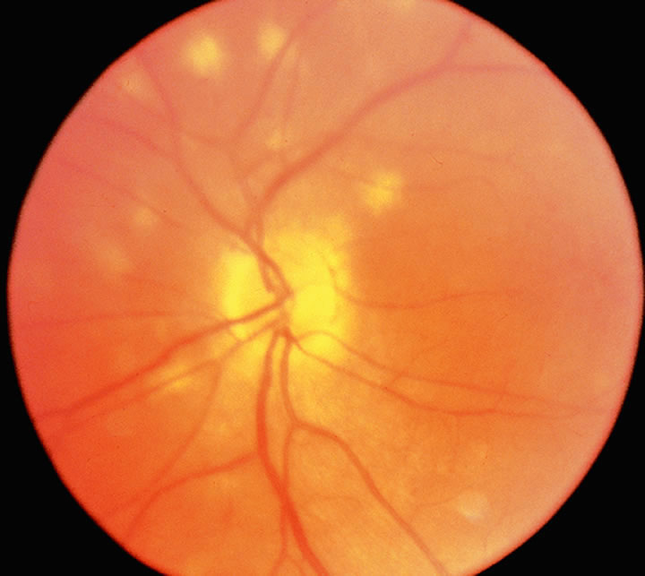
Reiter's Syndrome Reiter's syndrome is a postinfectious autoimmune disease that has strong associations with the HLA-B27 locus. The classic triad of polyarthritis, papillary conjunctivitis, and urethritis, as well as highly characteristic mucocutaneous lesions, are the most specific signs of Reiter's syndrome.66 Uveitis and iritis are also prominent ocular findings of this disease. Sexually transmitted chlamydial infection, or acute dysentery resulting from gram-negative organisms such as Klebsiella, Salmonella, Shigella, or Yersinia species often precedes the development of joint inflammation, hence the term “reactive arthritis.” Thirty-five percent of patients with sexually acquired reactive arthritis (SARA) following nongonococcal urethritis have conjunctivitis and other lesions characteristic of the full complex of Reiter's syndrome. There are a variety of theories to explain the role of HLA-B27 in the pathogenesis of reactive arthritis. It is likely that the features of HLA-B27 as an antigen-presenting molecule; the possible role of the free B27 heavy chains; and the ability to bind long extracellular peptides, as well as the single cysteine at position 67 in the peptide chain, may all play a role in the pathogenesis.113,79 Birdshot Chorioretinopathy Birdshot chorioretinopathy is a clinically distinct form of chronic posterior uveitis with bilateral, multiple, hypopigmented postequatorial retinal pigment epithelial (RPE) and choroidal inflammatory lesions (Fig. 4). This disease is often complicated by cystoid macular edema, posterior synechiae, cataract, and vitreitis. The association between birdshot chorioretinopathy and HLA-A29, which has been documented as high as 95.6% with a RR factor of 224.35, is probably the strongest established to date for any disease.81,82
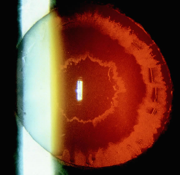
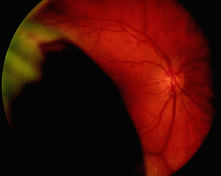
Presumed Ocular Histoplasmosis Syndrome (POHS) POHS is a triad of peripheral, punched-out chorioretinal atrophic lesions; peripapillary atrophy; and macular subretinal neovascular membrane formation specifically in the absence of vitreitis. It is strongly associated with skin test responsiveness to Ag from the filamentous fungus Histoplasma capsulatum, which is endemic to the Mississippi River valley. Certain individuals in endemic areas may have an increased susceptibility to POHS following exposure to the fungus, as well as an additional propensity to develop visually disabling macular lesions. HLA-B7 is strongly associated with disciform macular lesions83 but not with peripheral atrophic scars seen in POHS. HLA-DR2, on the other hand, is associated with both macular and peripheral scarring,84 which incriminates a genetic predisposition to macular neovascularization in this syndrome and apparently has a differing genetic risk than multifocal choroiditis and panuveitis.84 Vogt-Koyanagi-Harada (VKH) Disease VKH disease is a rare multisystem disease affecting the eye, inner ear, the skin, and the central nervous system.85 It is believed to be due to an autoimmune condition in which cytotoxic T cells target the melanocytes leading to uveal depigmentation, poliosis, and vitiligo. Patients present initially with iridocyclitis and choroiditis, occasionally severe enough to produce an exudative retinal detachment. Many of the initial studies on this syndrome demonstrating significant HLA associations have been performed on Japanese populations. The syndrome has been associated with HLA-Bw54 (a split of HLA-Bw22 also known as Bw22J), HLA-DwWa (a new D locus Ag), and HLA-DR4. This syndrome has been described in Caucasians, in whom these Ags do not appear to be associated with the disease process.84 VKH has been demonstrated to have combined allelic predisposition in which DQA1*0301 acts as the primary factor and HLA-DR4 acts as an additive factor in the development of the disease. There is also a negative association of DQB1*0604; it has been proposed that DQB1*0604 may provide protection.87 There have been some case reports of VKH syndrome following cutaneous injury, which suggests that this disorder may result from systemic sensitization to shared melanocytic Ags.88 Ethnicity is also a complicating factor with HLA association, because VKH is more common in persons with higher levels of skin pigmentation. In non-Japanese studies, HLA-DRB1*0405 has been associated with Brazilian patients,89 and DRB1*0101was associated with Mexican populations.89 Sympathetic Ophthalmia Sympathetic ophthalmia is a rare uveitic syndrome seen in patients who had a prior history of penetrating trauma. A Japanese study found these patients to have HLA associations similar to those found in VKH85,90,91 (see Table 2). Experimental autoimmune uveitis is an animal model of disease that has similar findings to sympathetic ophthalmia.92 Studies of this disease have demonstrated the possibility of retina S-Ag as a factor in this disease.93 Glaucomatocyclitic Crisis Also known as the Posner-Schlossman syndrome,94 glaucomatocyclitic crises are heralded by acute, unilateral attacks of often severe ocular hypertension associated with mild iridocyclitis. A strong association with HLA-Bw54, determined to not be related to linkage disequilibrium, has been described in Japanese patients with this syndrome.95 Pseudoexfoliation Patients with glaucoma resulting from capsular pseudoexfoliation were more likely found to be HLABw35 positive than the other study patients with chronic, open-angle glaucoma in a Scandinavian study (Fig. 5).96 This conclusion was not confirmed in a subsequent study on a similar population.97 In an Irish study, an HLA association with pseudoexfoliation is identified for 14 Ags. Eleven Ags (HLA-A1, -A33, -B8, -B47, -B51, -B53, -B57, -B62, -DR3, -DR12, and -DR13) are significantly more common in the pseudoexfoliation group, whereas 3 Ags (HLA-B12, -B17, and -DR2) are significantly less common.98 Once again, more extensive studies are needed to confirm the association.
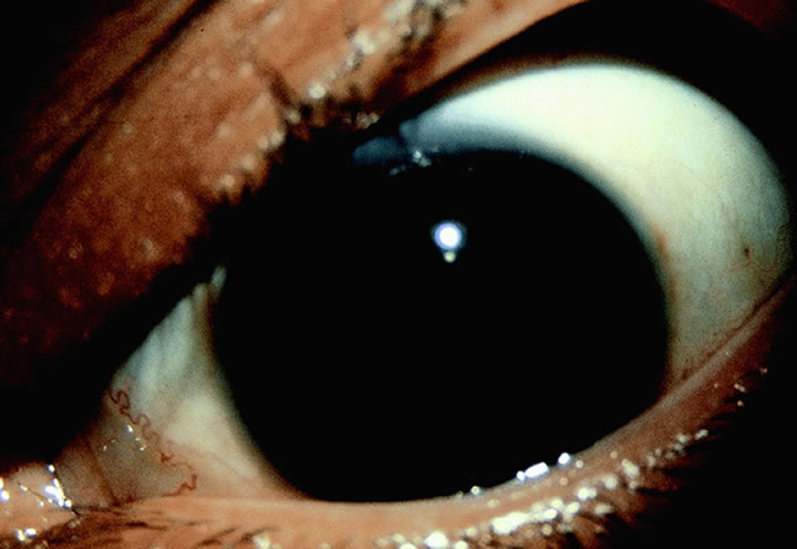
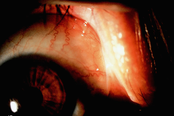
Behçet's Disease Behçet's disease is a multisystem vasculitic disorder with widespread clinical manifestations, including iritis, recurrent oral and genital ulcers, occlusive retinal vasculitis, arthritis, central nervous system involvement, and skin lesions (Fig. 6). An association of HLA-B5 was first described in Japanese patients in 1973.99 Later this was confirmed and further specified as an association with HLA-B51 and not of an associated with HLA-B52 (the two split products of HLA-B5). This association was found in Turkish, Jewish, English, and Mediterranean populations.100,101 Many of these populations are found along the ancient east and west passage known as the Silk Route that was used in medieval times to bring the material to Europe.102 The HLA association is less clear in Northwestern Europeans and Americans. Early studies in the 1970s did not show an association, but later findings found an increased frequency of HLA-B51 associated with those of Irish, Scottish, and German descent (far from the silk route).100 A separate study failed to find an association with those in an English clinic.103 Some authorities classify Behçet's disease into four subtypes: mucocutaneous, arthritic, necrologic, and ocular. HLA-B12 appears to be elevated in the mucocutaneous and arthritic groups, whereas HLA-B5 is increased in the ocular subgroup.104 About 70% of patients with Behçet's have ocular involvement,101 but there are no immunologic predictors indicating which patients will develop ocular disease. Ocular involvement may be the presenting sign in 20%. Refractory Behçet's disease may be associated with HLA-DQ genomic variations as detected by restriction endonuclease studies.103 Juvenile Rheumatoid Arthritis (JRA) Patients with pauciarticular JRA are particularly prone to uveitis, whereas those with Still'sdisease (systemic onset polyarthritis) and polyarticular rheumatoid factor positive JRA are usually spared.105 Still's disease is associated with HLABw35, whereas rheumatoid factor positive polyarticular JRA is associated with DR4. Further associations with DRB1*04 were observed in adults. DPB1*0201 was increased in the persistent pauciarticular group. DPB1*0402 was decreased in all pauciarticular groups with or without conversion.106 HLA-DR5 is associated with the early onset form of pauciarticular JRA seen most frequently in girls. Fifty-three percent of these children are likely to develop chronic bilateral ocular disease, band keratopathy, synechiae, and cataract and show positive antinuclear antibody (ANA) titers.67 Later onset pauciarticular JRA is more frequently seen in males and is a subset of HLA-B27 disease. Twenty-five percent of these patients develop uveitis. In poorly characterized JRA, the use of HLA typing for specific Ags may suggest the prognosis for ocular involvement. Nonuveitic Corneal and Conjunctival Disease with HLA Associations The first reports of association of keratoconus with HLA was by Gasset and colleagues107 who found one family in which a mother and her two children with severe keratoconus had HLA-B27 positivity; HLA-B27 negativity was found in the family members with no evidence of keratoconus. Keratoconus has been found to have HLA association in familial cases in Finland.108 More recently, the HLA-B27 Ag was demonstrated in the phenotype and increased incidence of haplotypes A2-B27 and A3-B35 were typical of patients with the progressive course of keratoconus, whereas in the acute form of keratoconus, there was an increased incidence of HLA-A1, -A19, -B7, and complete absence of HLA-A2.109 Other studies failed to demonstrate an association. More extensive and reliable studies are needed to determine the association of keratoconus and HLA. In Cogan's early descriptions of what has ultimately been called Cogan's syndrome, three of the four patients were found to have W-17 histocompatability Ag positivity.110 In a later study, HLA typing of 10 patients with well-documented Cogan's syndrome failed to support previous reports of an increased incidence of HLA-B17. Although HLA-A9 (Aw24), Bw35, and Cw4 appeared increased in frequency among patients, the small number of cases precluded attaching importance to the significance of these increased frequencies.111 Mooren's ulcer has been found to have an increased frequency of HLA-DR17 and -DQ2.112 An aberrant expression of HLA-DR Ag was found in keratoconjunctival epithelial cells and keratocytes in the corneal stroma biopsied from patients with Mooren's ulcer, implicating an excessive autoimmune reactivity as possibly the direct cause of Mooren's ulcer.113 Ocular cicatricial pemphigoid, an autoimmune mediated disorder (Fig. 7), was initially reported to be associated with HLA-B12, whereas later studies did not confirm this association. Other studies found that HLA-DR-4, DR-5, DQw3, B8, B35, and B49 have associations. With the use of RFLPs, HLA-DQB1*030 was confirmed in several studies to have an association.114 This association has led to further studies demonstrating that cell-mediated immunity, rather than antibody-mediated immunity may be a critical cause of the scarring in this disease.114
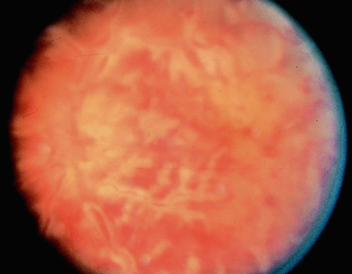
Stevens-Johnson syndrome has been associated with the DQB*0601 allele, with a RR of 7.2.115 HLA-Bw44 (a split from HLA-B12, which is of interest because of the association with ocular cicatricial pemphigoid) was also found to have an association with Stevens-Johnson syndrome.116 The HLA association may be one of several factors contributing to this autoimmune disease, because immune complexes have been demonstrated in the blood and immune globulins are deposited at the dermal-epidermal junction.115 Thygeson's superficial punctate keratopathy has been associated with HLA-DR3.117 Ophthalmic Diseases with Conflicting Reports of HLA Association As more information is gathered about HLA and their associations with disease, it becomes clearer that HLA polymorphisms appear to be responsible for genetic variations in the extent of the immune response seen in different individuals in response to different Ags. There have been many studies in which no MHC Ags could be linked to ophthalmic diseases. In the field of cataract, senile cataract has not been found to have any HLA association.118 Diabetes, mitochondrial myopathies, and uveitis are all associated with the formation of cataracts, but there has been no demonstrable HLA association with the cataract.119 Primary open-angle glaucoma (POAG) is familial in 20% of the cases; however, even with the considerable evidence of genetic factors in POAG, there have been inconsistent reports.120 Various studies demonstrate association with increased and decreased association with various HLA types; however, the associations with HLA-D and -DR have not yet been studied in detail.119,120 There have been no HLA associations on the A or B loci found for acute angle closure glaucoma.120–122 No association was found with pigment dispersion glaucoma,123,124 oral steroid-induced ocular hypertension and steroid-induced cataract, or chronic uveitis. There have been inconsistent reports of HLA associations with herpetic keratitis. HLA Antigens and Fundus Diseases Retinoblastoma (Rb) was found to have a possible HLA association in a small study in the 70s,125 whereas another study of 16 Rb patients did not confirm this.126 Interestingly, when a patient has Rb, it has been shown that the expression of Rb leads to modification of the chromatin environment of the HLA-DRA promoter independent of transcription.127 Eales's disease was recently demonstrated to have a positive disequilibrium of haplotypes A3-B44 and A11-B12;128 however, there was no association demonstrated in other studies. Choroidal malignant melanoma (Fig. 8) has been associated with HLA-Aw32129 and a loss of heterozygosity on chromosome 6,130 but a separate study of 514 patients with melanoma in other parts of the body and a study of 64 patients with uveal melanoma failed to reveal any significant association.131 No HLA association was demonstrated with central serous retinopathy,129 chorioretinitis, toxoplasmic retinochoroiditis, chronic juvenile iridocyclitis, age-related disciform macular degeneration, retinitis pigmentosa, or rhegmatogenous retinal detachment. Proliferative vitreoretinopathy (PVR) was found to have increased HLA-DR expression on RPE cells; this could play a role in triggering a local immune response in PVR.132 Sympathetic ophthalmia was found in an early study to have no HLA association,90 whereas in a Japanese study, a significant association was found with HLA-DR4 and DR5391; and in North American patients, an association with DR-4 was noted.85 Ophthalmic Diseases with Systemic Components and HLA Sarcoidosis has not been found to have a consistent HLA association.133 Diabetes mellitus has been evaluated closely since 1972 for HLA associations.134 There has been an increased risk of insulin-dependent diabetes mellitus (IDDM) in HLA-Dw3 or DRw3 positive patients. In retinopathy, a variety of HLA associations have been suggested including the risk for advanced retinopathy in HLA-B15 positive patients and more common severe retinopathy in patients with DR3-DQ2/DR4-DQ8 alleles.135 Temporal arteritis has been found to have an association with HLA-DRB1 *04 alleles.136–138 There is considerable evidence that Graves' disease is of autoimmune etiology, and there have been several studies suggesting a significant association between HLA-B8 and Graves' disease in Caucasian populations.21,22,120,139 Molecular mimicry appears to play a role in the pathogenesis. |
TRANSPLANTATION IMMUNOLOGY | |
Corneal transplantation has the lowest rejection rate among the solid tissue
allografts performed in humans. Greater than 90% of transplants
described as low risk survive for more than 2 years.140 The successful long-term preservation of a corneal graft is highly dependent
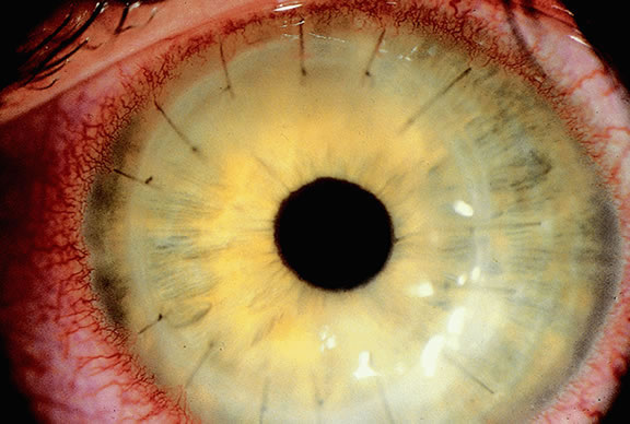
on the presence of host vascularization.3,140,141 Rejection reactions often begin as a line of infiltrating leukocytes (Khododoust's
line) at the site of maximal vascularization (Fig. 9). Other inflammatory foci such as loose sutures, suture removal, trauma, or
continuing inflammation (such as in chemical burns or previous graft
failure) leading to vascular dilation can also lead to acute rejection
reactions.3,142 This reflects the features of the cornea that contribute to its immune
privileged nature. The cornea lacks blood vessels, lymphatics, and bone
marrow-derived cells. The cells of the cornea secrete immunosuppressive
factors including interleukin-1 receptor antagonists and transforming
growth factor beta. These and other features explain the high success
rate of allograft survival in normal eyes.140 As early as 1976, studies evaluated the benefit of HLA matching in corneal
grafts, and most found no significant benefit to matching of HLA.32,40,55,61 However, there has been some evidence supporting that minor histocompatability (mHag) may
affect the graft-host compatibility.143,144 Other studies demonstrated that in high-risk patients, grafts fully matched
for HLA-A, -B, and -DR had a significant decrease in rejection.145 Suppressor T cells may be instrumental to preservation of corneal grafts,143 even following rejection reactions. Repeat grafts also have an increased
incidence of rejection, resulting from host sensitization, vascularization, and
increased vascular permeability following a permanent rejection. Unlike
renal allografts, corneal transplant rejections have not
been shown to be more frequent in multiparous patients or blood transfusion
recipients. Increased rejection rates have been associated with
eccentric grafts in which the foreign Ag is anatomically closer to limbal
vascular and lymphatic channels and with large grafts (grater than 8.5 mm
in diameter in the normal eye) in which a greater antigenic
load is placed in close proximity to the limbus.144,146 It would seem that the peripheral distribution of Langerhans' cells play
a role in eccentric and large-sized graft rejection. However, Stulting
and coworkers147 demonstrated that removing the donor epithelium, and hence the Langerhans' cells, does
not decrease the incidence of rejection. Donor tissue
younger than 2 years old, in addition to presenting technical difficulties, is
associated with increased rejection rates, possibly resulting
from enhanced Class II expression on donor epithelial cells. Patients
younger than the age of 20 also have an increased risk of corneal allograft
rejection, presumably because of the general vitality of their
overall immune responsiveness.141,148 There has been some convincing evidence that matching for HLA Ags improves
cornea graft survival in patients with highly vascularized corneas
or previous graft failures; this, however, continues to be under investigation
by the Collaborative Corneal Transplantation Study (CCTS).145,146,149
It is not entirely clear whether HLA matching is beneficial to the survival of uncomplicated, nonvascularized corneal transplants for conditions such as keratoconus or central corneal scarring. Very favorable rejection rates have also been attained in high-risk patients by obtaining donor tissue with HLA Ags to which the recipient was not sensitized. Previous sensitization is determined by a lymphocyte crossmatch study. Two disincentives to HLA matching is the high degree of polymorphism and the cost and the delay in time with traditional serology typing techniques. Studies evaluating HLA-matching for stem cell transplantation have demonstrated a possible benefit with HLA-matching. One patient with a complete HLA mismatch had poor epithelial healing and a trophic ulcer. Because the conjunctiva is a vascularized tissue, some groups have decided to use donors with at least a 50% HLA match.150 HLA expression is thought to be altered in the face of herpes infection, autoimmune reactions, and vascularization.151 This elevated levels of HLA may make these corneas more susceptible to attack by cytotoxic cells and hence graft rejection.152 However, the genetic susceptibility to corneal herpetic disease appears to be dependent on non-MHC loci.42 Graft rejection in patients with previous herpes infection has been shown to be associated with induction of Class I HLA-ABC and Class II HLA-DR Ags on cells in the donor button by a factor associated with cellular inflammation.153 COLLABORATIVE CORNEAL TRANSPLANTATION STUDY The CCTS is addressing the value of donor-recipient histocompatibility matching by pooling high-risk recipients and donor tissue through a nationwide network for donor selection and tissue distribution.154 This multicenter, randomized, double-masked clinical trial is assessing recipients for detectable lymphocytotoxic antibodies. Those who have antibodies will be assigned randomly to receive either a negatively or positively crossmatched cornea. Patients without detectable antibodies are assigned donor tissue with varying degrees of HLA matching. They found that there was no difference in highly matched (zero or one HLA-A, -B mismatch) or low matched (two or more mismatched transplants).3,40,148 In fact, an inverse correlation for HLA-DR was found, in which incompatible grafts had a superior survival rate compared with matched grafts.155 One confounding factor for the CCTS is the aggressive use of immunosuppressants leading to a high overall survival of the high-risk grafts (65% at 3 years).148 The CCTS group did find a significant survival advantage with blood group compatibility, which was previously believed to have little or no relevance. A non-CCTS, retrospective analysis found rejection in 1681 patients to be almost 14 times greater in recipients with HLA-AB and HLA-DR mismatched donor corneas.149 The diversity of experimental results and complexity of the HLA system confirms the need for more study of the benefit of HLA matching for corneal transplantation. |
ACKNOWLEDGMENTS |
The authors would like to thank John Troupe, Bruce Tonjes, Arlene Tonjes, and Christopher Muller for their editorial support on this project. In addition, the editors and authors wish to acknowledge the contributions of the previous authors of this chapter, John D. Shepard MD and Marvin R. Garvey MD. Some of their material has been adapted to this revision. |