Chapter 41
Normal Flora of the Human Conjunctiva and Eyelid
REGIS P. KOWALSKI and MELVIN I. ROAT
Main Menu   Table Of Contents

Search

FORMATION OF NORMAL FLORA
NORMAL BACTERIAL FLORA
TAXONOMY AND TECHNIQUES
NORMAL NONBACTERIAL FLORA
HOST FACTORS AFFECTING THE FLORA
SUMMARY
REFERENCES

Microorganisms are present on the normal human conjunctiva and eyelid without causing ocular disease. These isolates are either contaminants from the environment, spread to the eye from other areas of the body, or colonizers that thrive independently. Bacteria are the principle microbe isolated from the normal conjunctiva and eyelid. The types and numbers of bacteria are limited by human and bacterial defense mechanisms. Any deficiencies or shifts in these defenses may alter the flora. This chapter describes the microbial flora of the normal and compromised human conjunctiva and eyelid.
Back to Top
FORMATION OF NORMAL FLORA

COLONIZATION VERSUS RECURRENT CONTAMINATION

Microbes may be present on the eye as the result of two different processes. The first, colonization, results from the establishment of a stable, independent, virtually self-perpetuating community of microbes. The proliferation of the microbes is balanced by the host defenses, which results in a stable number of microbes being maintained over a long period without producing disease. The second mechanism, recurrent contamination, requires repeated introduction of microbes as host defenses continuously reduce the counts until the microbe is eliminated. When recurrent contamination is frequent or continuous, it results in a stable microbe population in which, as in colonization, the microbe count can remain stable over a long period. New microbes may be introduced from the environment, adjacent tissues, or from distant tissues by fomites (e.g., hand to eye ). Many of the microbes found on the eyelids have colonized the eyelids, and others are recurrently introduced from adjacent skin and to a lesser extent from the upper respiratory tract (the nose) and the upper gastrointestinal tract (the mouth). This is in contrast to the conjunctiva, where colonization is generally a minor component and continuous contamination from the eyelids is usually the main source of microbes.

MICROBE FACTORS

Microbial adherence is important for colonization of the eyelids but it is especially critical for colonization of the conjunctiva, where there are mechanisms to wash out and scrape the conjunctival surface. Bacteria are able to adhere to epithelial cells using several techniques. Bacterial cell wall-surface glycoprotein adhesins (invasins) attach to epithelial cell membrane-surface protein receptors (integrins).1 For example, the teichoic acid fraction of the Staphylococcus aureus cell wall is an adhesin to epithelial mucosal cells.1 There are other microbial cell wall components (microbial surface components recognizing adhesive matrix molecules) that facilitate microbial adhesion to epithelial cell surface ligands such as collagen, laminin, fibronectin, fibrinogen, vitronectin, heparin sulfate, thrombospondin, and elastin.1 Some gram-negative bacteria, such as Pseudomonas, Neisseria, Moraxella, and Haemophilus species, use pili (fimbriae) to adhere to epithelial cells.1 Biofilm (slime) production can promote adhesion and protect the bacteria from other host defenses.1

Microbes interact with other microbes in several ways. The receptor-attachment sites may be occupied by normal flora, thus limiting access to receptor-attachment sites by potential pathogens. Bacteria protect themselves from other bacteria by producing bacteriocins. These proteins are lethal to closely related bacteria. The metabolic end products of bacteria such as acetic and lactic acids also inhibit the growth of other bacteria.

HOST DEFENSES

The ocular surface is a moist, warm tissue with a high oxygen tension and an ample supply of nutrients. Without adequate host defenses, contaminating microbes would colonize, colonizing microbes would invade, and invading microbes would infect.

The eye has several anatomic, physical, biochemical, and immunologic defense mechanisms against microbes. Anatomic and physical defense mechanisms include:

  An intact epithelium, which is important in preventing most microbes from invading the conjunctiva and eyelid.
  Eyelashes, which filter dust and contaminated particles to prevent entrance to the ocular surface.
  The blink reflex, which is initiated when an object or insect touches the eyelashes, preventing access to the ocular surface.
  The blinking action of the eyelids, which sweeps debris and organisms from the ocular surface to the lacrimal puncta, where it is sucked into the lacrimal duct and then into the nose.
  Normal and excessive tearing, which washes bacteria and debris from the conjunctiva.
  Conjunctival and eyelid skin epithelial cells, which are frequently and continuously desquamated. This prevents invasion by microbes and leads to shedding of microbes adherent to the superficial layer.

Biochemical defenses include products produced by the eyelids, conjunctiva, and lacrimal glands. Some bacteria and fungi are vulnerable to unsaturated and long-chained fatty acids produced by thehuman meibomian glands.2,3 The lipids also produce an acidic environment, which selects against some types of bacteria.1 The tear film contains many soluble components to limit microbial flora. Lysozyme is lethal to Micrococcus species and other bacteria (in the presence of specific immunoglobulins and complement). Metal-chelating proteins such as lactoferrin bind iron, which interferes with bacterial iron uptake and metabolism. This interference can decrease bacterial virulence and allows some bacteria to be more susceptible to other antimicrobial tear components. β-Lysin acts on the cytoplasmic membranes of bacteria, leading to lysis.

The tears also contain complement and immunoglobulins. The components of complement that are found in tears represent two separate pathways that are cascades of reactions that lead to a microbiological defense.4,5 Complement, when activated leads to chemotaxis of effector cells; opsonization, resulting in enhanced phagocytosis of bacteria and protozoa; production of an anaphylatoxin; and lysis of virus-infected cells, bacteria, and protozoa. Immunoglobulins act against bacteria in several modes. IgA prevents bacteria from adhering to the conjunctival epithelium, activates the alternate complement pathway, and neutralizes viruses and toxins. IgG binds to microbes resulting in opsonization (independent of complement), with subsequent enhanced phagocytosis of bacteria and protozoa; antibody-dependent cell-mediated cytotoxicity of bacteria, virus-infected cells, and protozoa; neutralization of viruses and protozoa; and activation of the classic complement pathway, with all of the resulting effects.The cellular elements of the immune system are also active against microbes. Polymorphonuclear leukocytes can phagocytose bacteria, fungus, and protozoa. The process is facilitated by the binding of opsonizing antibodies or complement. Macrophages can act as antigen-presenting cells, phagocytose bacteria and protozoa, and have a major role in defense against fungi, mycobacteria, and some protozoa when lymphokines are activated. Cytotoxic T cells and natural killer cells are capable of recognizing and killing virus-infected cells, independent of immunoglobulins. Killer cells, an effector cell of antibody-dependent cell-mediated cytotoxicity, are active against bacteria, virus-infected cells, and protozoa. Mast cells degranulate when specific IgE on their surface bind to antigens on a protozoa. The released effector molecules include histamine (leading to excess tearing, in addition to other effects) and chemotactic factors, among others. Eosinophils degranulate when they contact IgE-coated protozoa, releasing effector molecules such as major basic protein, which is highly toxic to protozoa.

The conjunctiva and eyelid have a rich blood supply, which allows an abundant response by the humoral and cellular elements of the immune system. The conjunctiva and eyelid also have an excellent lymphatic network and the local conjunctiva-associated lymphoid tissue, both of which are important in the afferent arm of the immune response.

A balance of the bacterial and human defense mechanisms dictates the normal microbial flora of the human conjunctiva and eyelid. Infection results when there is an imbalance between 1) proliferation of the microbes and the invasive potential of the microbes, and 2) the defenses by other microbes that make up the flora and the host. Because an intact stratified squamous epithelium is not easily invaded by most pathogens, the first step leading to infection is attachment of microbes to the epithelial surface. Adhesion occurs more to damaged than nondamaged epithelium. Microbial attachment is not necessarily followed by epithelial cell penetration. Some microbes infect only the epithelial surface, whereas others invade the epithelial cell or subepithelial tissue.

The ocular surface and host defense differ for the closed eye during sleep, compared with the open eye. During sleep, the ocular surface is dryer because of a lower volume of tear production, which may be due to reduced light and sensory stimulation of the lacrimal gland,6 and the conjunctival vessels are dilated. A subclinical inflammatory state with an influx of polymorphonuclear cells7–9 exists due to an increase of fibronectin10 from the blood vessels, interleukin-8 from the tears,11 and the conversion of complement C3 to C3c.7 The concentration of secretory IgA is 40 times greater in the closed eye than the open.8 The closed eye also results in a decrease of oxygen to the conjunctiva and ocular surface, which alters the epithelium and opens receptor sites to bacterial attachment.12

Back to Top
NORMAL BACTERIAL FLORA

CONJUNCTIVA

The human conjunctiva is first exposed to microbes when an infant passes through the birth canal.13,14 Right after birth, the eye is exposed to environmental sources of bacteria such as air, contaminated material, and people. The flora obtained from the birth canal are predominantly aerobic and anaerobic bacteria (Table 1). The most common aerobic bacteria isolated immediately after birth from the conjunctiva are Staphylococcus aureus (coagulase-negative), diphtheroids (Corynebacterium species), Streptococcus species, Enterococcus species, and Escherichia coli. Propionibacterium acnes, Bifidobacterium, and Bacteroides species are isolated anaerobically from the newborn conjunctiva after vaginal delivery. Lactobacillus species, a microaerophilic bacteria, can also contaminate the conjunctiva from the vaginal tract. Other anaerobes and aerobes have been isolated less frequently (see Table 1). The conjunctiva is sterile in 20% to 87% of infants born vaginally.15 The conjunctiva cultured after cesarean section is sterile in 80% to 95% of infants.15,16 If cesarean section is delayed for more than 3 hours after the sac is ruptured, conjunctivas are sterile in 45% of newborns.


TABLE 41-1. Bacterial Flora of Normal Human Conjunctiva from Birth through Various Age Groups


LocationCaliforniaKoreaCaliforniaKoreaWashingtonCaliforniaCaliforniaGeorgiaHawaii
InvestigatorBrook, 197913Lee, 198916Brook, 197913Lee, 198916Weiss, 199260Singer, 198817Singer, 198817McNatt, 197861Brown, 197862
Source of specimenNewbornsNewbornsNewbornsNewbornsChildrenPresurgicalPresurgicalVolunteersNursing home
AgeAfter birthAfter birth48 hr48 hr4 mo to 12 yr<17 yr>17 yr22–60 yr26–99 yr
Number of eyes47186481869147 less than182184118
Bacterial activity (no. isolates)         
 No growth 136 (73%)-112 (60%)23 (25%)11 (23%)39 (21%)72 (39%)16 (14%)
 Coagulase-negative Staphylococcus10 (21%)28 (15%)17 (35%)40 (22%)9 (10%)14 (30%)108 (59%)43 (23%)57 (48%)
 Staphylococcus aureus06 (3%)013 (7%)2 (2%)3 (6%)7 (4%)1 (0.5%)14 (12%)
 Diphtheroids/Corynebacterium8 (17%)-9 (18%)-4 (4%)17 (36%)82 (45%)19 (10%)35 (30%)
 Streptococcus viridans group9 (19%)-9 (18%)------
 Streptococcus species----1 (1%)7 (15%)4 (2%)4 (2%)1 (1%)
 Enterococcus species1 (2%)-1 (2%)-1 (1%)---5 (4%)
 Micrococcus species2 (4%)-8 (17%)--0 <2 (1%)6 (3%)-
 Bacillus species5 (11%)-1 (2%)--en>1 (2%)---
 Haemophilus species7 (15%)-2 (4%)--05 (3%)--
 Escherichia coli1 (2%)26 (14%)027 (15%)----1 (1%)
 Klebsiella species---4 (2%)----1 (1%)
 Proteus species------2 (1%)-14 (12%)
 Propionibacterium acnes6/27 (22%)-10/18 (55%)--6 (13%)55 (30%)72 (39%)-
 Bacteroides species7/27 (26%)-0/18------
 Peptostreptococcus species9/27 (33%)-4/18 (22%)--01 (0.5%)5 (3%)-
 Lactobacillus species3/47 (6%)-1/48 (2%)------
 Eubacterium species1/27 (4%)-2/18 (11%)------
 Clostridium species2/27 (7%)-2/18 (11%)------


During the first several days of life, the infant's own cutaneous, respiratory tract, gastrointestinal flora, and the flora of the surrounding environment are important new sources of bacteria for the conjunctiva. After birth, before prophylaxis, there is a dramatic increase in the frequency of culture-positive conjunctivas, from as few as 13% of newborns to 98% of 3- to 5-day-old infants. Bacteria isolated include Staphylococcus species, Streptococcus species or Moraxella (Branhamella) catarrhalis.15

The literature (see Table 1) reports that from birth to old age, the conjunctiva is culture-negative for bacteria in 73% to 14% of patients. The frequency of isolating bacteria from the conjunctiva increases with age (Fig. 1). Coagulase-negative staphylococcus, diphtheroids (Corynebacterium species), P acnes, and S aureus are the most frequently isolated bacteria from the normal conjunctiva. Generally, the density of isolation is limited to fewer than 10 colonies on culture, compared with the confluent growth of infection. Streptococcus species are isolated more commonly from children than from adults. P acnes, diphtheroids, and gram-negative bacteria are isolated more frequent in adults.17,18 Less commonly, other gram-positive and gram-negative bacteria are isolated from the conjunctiva (Table 2). These isolates are probably transitory residents passed to the eye from other parts of the body and the environment by the adjacent skin or by the hands.

Fig. 1. Distribution of ocular bacterial isolates from the normal conjunctivas of newborns (Brook I, Barrett CT, Brinkman CR et al: Aerobic and anaerobic bacterial flora of the maternal cervix and newborn gastric fluid and conjunctiva: A prospective study. Pediatrics 63:451, 1979; Lee PW, Jun AK, Cho BC. A study of microbial flora of conjunctival sac in newborns. Korean J Ophthalmol 3:38, 1989), children (Weiss A, Brinser JH, Nazar-Stewart: Acute conjunctivitis in childhood. J Pediatr 122:10, 1993; Singer TR, Isenberg SJ, Apt L: Conjunctival anaerobic and aerobic bacterial flora in paediatric versus adult subjects. Br J Ophthalmol 72:448, 1988) and adults (McNatt J, Allen SD, Wilson LA et al: Anaerobic flora of the normal human conjunctival sac. Arch Ophthalmol 96:1448, 1978; Singer TR, Isenberg SJ, Apt L: Conjunctival anaerobic and aerobic bacterial flora in paediatric versus adult subjects. Br J Ophthalmol 72:448, 1988; Boes DA, Lindquist TD, Fritsche TR et al: Effects of povidone-iodine chemical preparation and saline irrigation on the perilimbal flora. Ophthalmology 99:1569, 1992.)


TABLE 41-2. Normal Flora of the Normal Human Conjunctiva Before Prophylaxis and Surgery


LocationWashingtonPolandHungaryEnglandSaudi ArabiaGreeceRussia
InvestigatorBoes, 199163Kecik, 199564Kovacs, 199465Bell/Walker, 86–8866,67Taylor, 198868Dereklis, 199469Karanadze, 198430
Number of eyes100236520017440504927
Bacterial activity (no. isolates)       
 No growth25 (25%)894 (38%)132 (66%)109 (63%)16 (40%)17 (34%)1166 (24%)
 Coagulase-negative Staphylococcus59 (59%)1152 (49%)43 (22%)74 (43%)22 (55%)17 (34%)1372 (28%)
 Staphylococcus aureus8 (8%)259 (11%)3 (2%)7 (4%)1 (3%)7 (14%)203 (4%)
 Diphtheroids/Corynebacterium2 (2%)10 (0.4%)1 (0.5%)6 (3%)4 (10%)2 (4%)2138 (43%)
 Streptococcus viridans group4 (4%)-1 (0.5%)-1 (3%)4 (8%)-
 Haemophilus species2 (2%)--2 (1%)---
 Enterococcus species1 (1%)-2 (1%)--2 (4%)8 (0.1%)
 Streptococcus species1 (1%)22 (1%)-1 (0.6%)2 (5%)-36 (0.7%)
 Streptococcus pneumoniae------80 (2%)
 Micrococcus species0-14 (7%)----
 Bacillus species---1 (0.6%)--46 (1%)
 Escherichia coli05 (0.2%)1 (0.5%)---100 (2%)
 Proteus species011 (0.4%)2 (1%)1 (0.6%)1 (3%)1 (2%)4 (0.1%)
 Pseudomonas species07 (0.3%)-1 (0.6%)---
 Citrobacter species0-3 (3%)1 (0.6%)1 (3%)--
 Klebsiella species1 (1%)5 (0.2%)-----
 Propionibacterium acnes6 (6%)------
 Peptostreptococcus species3 (3%)------


Locather-Khorazo and Seegal14 reported that the bacterial flora did not differ in the eyes of healthy males and females, nor between the right and left eye. There were also no significant fluctuations during different seasons and no changes in the recovery rate of Staphylococcus species and diphtheroids on a yearly basis. The bacterial flora were the same for the conjunctiva and eyelid and frequently a mixture of Staphylococcus species and diphtheroids (Fig. 2). When comparing the bacterial flora of the conjunctiva with subjects from different countries, we found that the types of bacteria isolated are similar, although the frequency varies.

Fig. 2. The growth of coagulase-negative staphylococcus and diphtheroids on 5% sheep blood agar with inserts of gram-stained morphologies. The long white arrows point to the broken-line circle containing the diphtheroid colonies and the insert of gram-positive pleomorphic rods that are characteristic of diphtheroids. The short white arrows point to the larger coagulase-negative staphylococcus colony and the insert of gram-positive cocci. (Photographed by Phyllis Kaufman, University of Pittsburgh Medical Center, Pittsburgh, PA.)

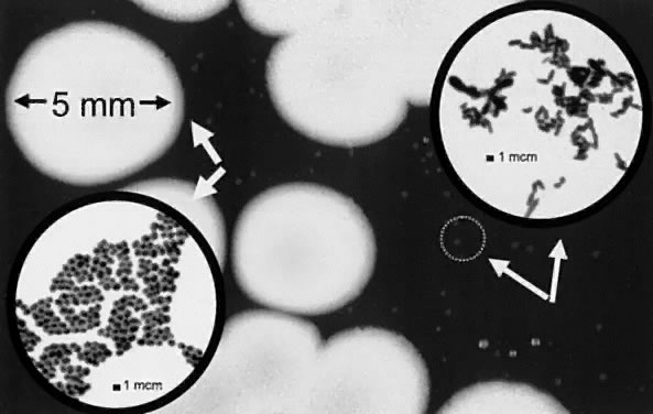
Table 2 details the bacterial flora isolated from human conjunctivas before surgery in selected parts of the world. The conjunctiva was 25% to 66% culture-negative. The conjunctivas were predominantly positive for coagulase-negative staphylococcus (22% to 59%); diphtheroids (Corynebacterium species, 2% to 43%); and S aureus (2% to 14%). Gram-negative bacteria on the conjunctiva were isolated more frequently in culture from eastern European countries. The isolation of gram-negative bacteria is generally limited to a few colonies on culture. Tomar and coworkers19 had reported a 6% recovery of Pseudomonas aeruginosa from the normal conjunctiva in India.

EYELID

The bacterial flora of the eyelid are similar to the bacterial flora of the skin. It is normal that a higher density of bacteria is isolated from the eyelid than from the conjunctiva. The normal human eyelid is always culture-positive for bacteria, and the bacterial types and quantities are random over time. Table 3 details the bacterial flora of the eyelid from several geographic locations. The types of bacteria are similar to those isolated from the conjunctiva. Coagulase-negative staphylococcus (575 to 100%) is the predominant bacterial isolate and generally exhibits heavy growth on culture. P acnes (71% to 74%), diphtheroids (8% to 45%), S aureus (12% to 16%), and Streptococcus viridans group (15%, children) are also frequently isolated with less density. A variety of gram-negative bacteria can also be isolated but less frequently and presenting with a few colonies.


TABLE 41-3. Flora of the Normal Human Eyelids


LocationWashingtonFloridaOklahomaSaudi ArabiaIreland
InvestigatorWeiss, 199260Groden, 199123Au, 199329Taylor, 198868Doyle, 199570
Source of specimenChildrenPatientsVolunteersPresurgical*Presurgical
Number of eyes91160984017
Bacterial Activity (no. isolates)     
 No growth00000
 Coagulase-negative Staphylococcus52 (57%)140 (88%)88 (90%)38 (95%)17 (100%)
 Staphylococcus aureus11 (12%)25 (16%)----4 (10%)
 Diphtheroids/Corynebacterium22 (24%)72 (45%)8 (8%)5 (13%)1 (6$)
 Streptococcus viridans group14 (15%)--------
 Enterococcus species3 (3%)--------
 Streptococcus species----1 (1%)--1 (3%)
 Proteus species----1 (1%)----
 Acinetobacter species4 (4%)7 (4%)------
 Propionibacterium acnes--118 (74%)----12 (71%)

*Before prophylaxis.


Back to Top
TAXONOMY AND TECHNIQUES

DIPHTHEROIDS

The term “diphtheroids” is a common reference to gram-positive pleomorphic rods that are isolated frequently on routine culture medium from the human conjunctiva and eyelids.20,21 Most organisms in this group exhibit various morphologies that include clubbing, branching, coccoid forms, and palisading. The pleomorphic shapes observed on gram staining have been referred to as “Chinese letter writing” (see Fig. 2). This group includes 32 genera of bacteria that are generally aerobic and non-spore-forming but can be anaerobic. Corynebacterium and Propionibacterium are the most frequent bacterial isolates from the conjunctiva and eyelid but other genera may be present. Diphtheroids are generally identified by gram staining morphology and are rarely further identified with laboratory biochemical tests.

The most common species of Corynebacterium isolated from the human conjunctiva is Pseudodiphtheria (C hofmannii) followed by C xerosis, C renale, and C mycetoides.22 Organisms in the diphtheroid group are often reclassified into other and new genera. Past Corynebacterium isolates from the conjunctiva have been renamed Arcanobacterium haemolyticum and Actinomyces pyogenes.

P acnes is a diphtheroid commonly isolated from the eyelid and occasionally the conjunctiva. P acnes is generally recognized as an obligate anaerobe but many isolates grow aerobically after the first passage from anaerobic medium. Many strains of P acnes grow from eye cultures on standard medium (especially chocolate agar), given 5 to 7 days incubation at 37°C in a CO2 incubator. Enriched thioglycollate is an excellent medium for isolating P acnes. P acnes and other diphtheroids may act as adjuvants in blepharitis and other ocular immunologic diseases.23

STAPHYLOCOCCUS

Staphylococcus species are gram-positive bacteria that appear as single, pairs, short chains, and grape-like clusters of cocci (see Fig. 2).24 Coagulase is a cell-associated clumping factor used for staphylococcal-species differentiation.The genus Staphylococcus is comprised of 27 species that are grouped as coagulase-positive (three species) and coagulase-negative (24 species). Staphylococcus species produce catalase and may produce acid on mannitol salt agar. Staphylococcus species are easily cultured on standard medium such as 5% sheep blood agar at 37°C, with or without a CO2 atmosphere, and colonies may appear with yellow-orange-gold pigmentation. S aureus is coagulase-positive, produces acid on mannitol salt agar, and generally appears as golden colonies on blood agar. More than 95% of S aureus produce protein A, which may be cell-associated or extracellular. Detection of protein A using rapid latex tests provides identification of S aureus. Some strains of S aureus may not produce acid on mannitol salt agar.

Coagulase-negative staphylococcus are the chief bacterial isolates from the normal eye, especially the eyelid. These isolates may or may not produce acid on mannitol. Staphylococcus epidermidis does not produce acid on mannitol and appears as whitish colonies on blood agar.24,25

S epidermidis (53% to 87%) is the most common coagulase-negative staphylococcus found in the normal conjunctiva and eyelid. Other coagulase-negative staphylococci found are S capitis (2% to 20%), S simulans (6% to 13%), S hominis (2.1% to 5%), S haemolyticus (4.6% to 6.9%), S xylosus (5.3%), S warneri (1% to 8.3%), S lugdunensis (2.6%), S cohnii (1.3% to 1.4%), S auricularis (0.6%), S caprae (0.6%), S saprophyticus (0.6%), and S sciuri (0.3%).26–29

GRAM-NEGATIVE BACTERIA

Gram-negative rods isolated from the normal conjunctiva and eyelid are easily grown on standard culture media. These are grouped by glucose fermentation and the oxidase reaction. Escherichia coli; Proteus (mirabilis, vulgaris, penneri); Klebsiella (oxytoca, ozaenae, pneumoniae); Serratia (marcescens, liquefaciens, rubidaea); Citrobacter (diversus, freundii); and Morganella (morganii) are oxidase-negative, glucose fermenters and are facultative anaerobes (grow readily with or without the presence of oxygen). Acinetobacter (lwoffi, calcoaceticus, baumanii) is an oxidase-negative, glucose nonfermenter. Moraxella (lacunata, osloensis) and Pseudomonas (aeruginosa) are examples of oxidasepositive glucose nonfermenters. These organisms require oxygen and do not grow well under anaerobic conditions.

Neisseria species (other than gonorrhoeae and meningitidis) and Moraxella (Branhamella) catarrhalis are examples of oxidase-positive gram-negative cocci. Haemophilus (influenzae) are oxidase-positive gram-negative coccobacilli that can be more fastidious with special nutrient requirements.

OTHER GRAM-POSITIVE BACTERIA

Gram-positive cocci such as Micrococcus species and Streptococcus species have been isolated from the healthy conjunctiva and eyelid. Micrococcus species appears microscopically as a group of four cocci (tetrads) on gram staining and yellowish colonies on culture media. Micrococcus species produces catalase, which separates it from the Streptococcus species. The normal conjunctiva is probably not colonized but contaminated with Micrococcus species from the eyelid and adjacent skin because the lysozyme in the tears is effective in eradicating this bacteria. A high incidence of Micrococcus species in the conjunctiva may indicate a deficiency in the production of lysozyme in the tears. There are several species of Micrococcus that are not normally pathogenic. Micrococci species are identified from gram staining and colony presentation on culture.

Streptococcus species isolated from the normal conjunctiva are generally of the viridans group. S viridans is catalase-negative, and colonies on blood agar are surrounded with a greenish halo called alpha-hemolysis. Other normal flora, Streptococcus species appear with beta-hemolysis (a clear halo) around the colonies, whereas some species demonstrate no hemolysis (no halo; gamma-hemolysis). Enterococcus faecalis, another infrequent isolate from the healthy eye, produces low-levels of catalase, is coccoid in appearance on gram staining, and demonstrates no hemolysis on blood agar.

The most common gram-positive rod isolated from the normal eye is Bacillus species. Bacillus species is ubiquitous, aerobic or facultatively anaerobic, spore-forming, gram-positive or variable, and may appear as large rods on gram staining. There are numerous species of Bacillus that are cumbersome to distinguish in the laboratory by ordinary biochemicals. Bacillus cereus is the most notable eye isolate that can be isolated normally but can be a severe opportunistic pathogen.

Back to Top
NORMAL NONBACTERIAL FLORA

FUNGI

Fungi, which include both yeasts and molds, are not normal colonizers of the human conjunctiva. Fungi are more often transient isolates of random contamination from the air and environment. Four independent studies from four distinct geographic locations reported an incidence of 1.3% to 13%.30–34 Ando and Takatori31 reported that 97 of 103 positive cultures (94%) yielded only one colony and the same fungus was seldom re-isolated from the same eye. The most frequent yeast isolated from the conjunctiva was Candida albicans. Aspergillus, Penicillium, Fusarium, Cladosporium, and Alternaria species were the principle isolated molds. There are no reports of the eyelids harboring more fungi compared with the conjunctiva. It is reasonable to conclude that the conjunctiva and eyelid may be contaminated by many airborne fungal contaminants.

The compromised eye may harbor more fungi in the conjunctiva than the normal conjunctiva. Satya and Tyagi32 reported that the incidence of fungi in an eye with clinical disease (conjunctivitis, keratitis, trachoma) was 24%. Nema and associates33 reported that the incidence of fungal isolates in trachomatous conjunctiva was also 24.76%. Ando and colleagues reported that 32 of 184 (17.4%) diseased eyes yielded fungi.31 Liotet and coworkers34 also indicated an increased incidence of fungi isolated from the conjunctiva of patients with immunity deficiencies.

Pityrosporon orbiculare (also called Malassezia furfur) is often observed on giemsa-stained smears from conjunctiva scrapings. These yeast-like organisms thrive in high-lipid areas such as the eyelid margin. These organisms require special medium for isolation and may have a pathogenic role in the seborrheic patient.

VIRUS

Viruses are not normal inhabitants of the human conjunctiva and eyelids because of their intracellular parasitic nature. Herpes simplex can sometimes be isolated from the conjunctiva of asymptomatic individuals.35 The virus is reactivated from its latent stage in the trigeminal ganglion and then shed into the conjunctival cul-de-sac. This recovery is transient and may be triggered by some stimuli of activation. In a study of human immunodeficiency virus(HIV)-positive and -negative individuals,36 normal conjunctivas were negative for herpes simplex virus DNA using the polymerase chain reaction. The same study detected Epstein-Barr virus DNA from 47% of HIV-negative normal conjunctivas. The lacrimal gland is believed to be a source of Epstein-Barr virus replication and nonpathogenic infection.37,38

PROTOZOA AND ARTHROPODS

Protozoans such as Acanthamoebae are not normal inhabitants of the human conjunctiva. Larkin and Easty39 reported that Acanthamoebae may use commensal bacteria from the conjunctiva and eyelid as a nutrient source. Stopak and associates40 demonstrated that cellular components of the cornea and conjunctiva could also provide the necessary nutrition for Acanthamoebae survival. Acanthamoebae is a ubiquitous protozoa found in water and soil. It could easily contaminate the eye but not remain and colonize the eye without causing disease.

Demodex folliculorum and bevis are arthropods that inhabit the hair follicles of the eyelids.1,41 These eight-puggy-legged cigar-shaped organisms are not culturable on standard media and can be identified only by observing clipped eyelashes under a light microscope. Although all age groups can harbor Demodex, the incidence in older patients may be 100% of the population.

Back to Top
HOST FACTORS AFFECTING THE FLORA

CONTACT LENSES

The types of organisms in the conjunctival flora of daily wear contact lens wearers differs somewhat from that of non-contact lens wearers, with studies indicating an increase in the number of species,42 an increase in the incidence of gram-negative types,43–45 and an increase in the number of bacterial organisms.42,46 In addition, some studies found a decrease in the number of positive cultures43,45,47 due to a decrease in the frequency of coagulase-negative staphylococcus isolated. Fleiszig43 reported an increase in the number of potentially pathogenic bacteria from the conjunctiva but these bacterial species are generally not implicated in producing keratitis. These bacteria were isolated more often from individuals using nonperoxidase disinfection systems. The increase in gram-negative isolation may be from contaminated solutions and contact lens cases and poor technique and cleanliness of some wearers.Larkin and Leeming42 reported no quantitative differences in the normal flora of wearers who used either soft or hard gas-permeable lenses. Fleiszig and Efron44 found no differences in the distribution of microorganisms.

IMMUNOSUPPRESSED HOST

The reports on the conjunctival flora of the HIV-positive patients indicate that the predominant bacterial organism is coagulase-negative staphylococcus. Gritz and colleagues48 compared the flora of 40 acquired immunodeficiency syndrome (AIDS) patients with 42 HIV-negative in a gender- and age-matched study and found no qualitative or quantitative differences in the conjunctiva or eyelid flora. Comerie-Smith and associates49 reported that HIV patients had decreased levels of lactoferrin and increased bacterial counts, compared with normal controls. Campos and coworkers50 reported that anaerobes, Clostridia, and Actinomyces species were isolated more frequently in the conjunctivas of HIV-positive patients compared with normal subjects. Gumbel and associates51 described a difference in the conjunctiva flora of out-patients and nosocomial AIDS patients. Out-patients were generally culture-positive for Staphylococcus epidermidis (36.6%) and although there was no conjunctival infection, nosocomial patients were found to have a normal incidence of Staphylococcus epidermidis (18.8%) and an increased incidence of S aureus (25%), C albicans (25%), and P aeruginosa (8.3%). This may indicate that HIV-positive patients are less proficient at clearing contaminants from the conjunctiva.

The conjunctivas of patients receiving immunosuppressant drugs have a greater variety of bacterial flora than normal controls. Patients receiving immunosuppressant drugs and normal controls had similar numbers of coagulase-negative staphylococcus (72% versus 74%), S aureus (3% versus 5%), diphtheroids (11% versus 16%), and S viridans (2% versus 5%). Although normal controls grew no other bacteria, the patients receiving immunosuppressant drugs grew many other gram-positive and gram-negative bacteria, such as Enterobacter species, Haemophilus influenzae, and Streptococcus group D. Both those receiving immunosuppressant drugs and normal controls grew similar numbers of fungi (3% versus 4%).52 The percentages of positive cultures and tear lysozyme levels are also equivalent. No difference was found in the ocular flora of patients with primary immunodeficiency syndromes (e.g., ataxia telangiectasia, chronic granulomatous disease, and hypogammaglobulinemia) compared with normal controls. In the same study, cultures for herpes simplex virus and Chlamydia species were negative in patients with primary immunodeficiency syndromes. More blepharitis and conjunctivitis are present in immunodeficient patients but the bacterial and fungal flora are no different than those found in normal controls.53

MISCELLANEOUS CONDITIONS

The bacterial flora from the anophthalmic conjunctiva are similar to those from the normal conjunctiva. The aerobic bacterial isolates consist of coagulase-negative staphylococcus, diphtheroids, Micrococcus species, S aureus, S viridans group, and a few gram-negative bacteria such as Klebsiella, Serratia, and Acinetobacter species, and P aeruginosa.54 The anaerobic bacteria isolated are Propionibacterium, Veillonella, and Peptostreptococcus species.50 The anophthalmic cavity can be easily contaminated by other gram-positive and gram-negative bacteria from the prosthesis and adjacent eyelid or skin.

The conjunctival flora from patients with severe thermal burns shift from coagulase-negative staphylococci to Staphylococcus aureus starting at day 5 to 6 postburn.55 Also at that time, diphtheroids diminish from the conjunctival flora and gram-negative bacteria that were not present on initial culture are detected. Examples include Enterobacter cloacae (18%), Klebsiella pneumoniae (15%), and especially P aeruginosa (34%). By serial culturing of other sites of the body and using phage typing, Pranhus55 determined that changes in the flora of the eye occurred simultaneously with changes in the flora of other sites. This supports the idea that many of the flora in the eye result from continual contamination and are not the result of colonization. Even though more pathogenic bacterial isolates are observed and many patients had corneal epithelial defects, most of these patients do not develop serious eye infections.

The conjunctival flora of leprosy patients without active ocular infection contain more pathogenic bacteria compared with normal controls.56 There is an increased incidence of positive cultures (98%). Of note were S aureus (34%) and gram-negative rods such P aeruginosa and Proteus mirabilis. Fungi (most common, Aspergillus, Alternaria, and Phialophora species) are also isolated from 79% of eyes.

The chronically inflamed conjunctiva is reported to carry more S aureus and gram-negative bacteria.57

The manipulation of the eye by routine examination, tonometry, and irrigation can temporarily change the bacteria isolated from the conjunctiva. Bacterial shifts from germ-free to increased numbers and isolation of potential pathogens can be observed. This is probably due to peripheral contamination.58,59

Back to Top
SUMMARY
The microbial flora isolated from the normal conjunctiva are predominantly bacterial, with a rare isolation of fungi. Virus and protozoa are not normally isolated from the conjunctiva. Because the conjunctiva is frequently culture-negative and bacterial flora is often identical to that of the eyelid and skin indicates that the bacteria isolated are likely present because of continual contamination than colonization. The most common bacteria isolated from the conjunctiva are small numbers of coagulase-negative staphylococcus and diphtheroids. The eyelid is heavily colonized with coagulase-negative staphylococcus and diphtheroids (including P acnes).

Contact lens, HIV-positive, and immunosuppressed patients have similar bacterial flora to that of normal controls but minor differences have been reported. Patients with chronic ocular disease present with more pathogenic bacteria such as S aureus, P aeruginosa, and other gram-negative bacteria. The shift in pathogens is not associated with an increase in the incidence of infectious conjunctival and eyelid disease. The normal flora of all individuals (normal or ill) appear to be an extension of the flora from other areas of the body.

Back to Top
REFERENCES

1. Osata MS: Normal ocular flora. I: Pepose JS, Holland GN, Wilhelmus KR (eds): Ocular Infection and Immunity, p 191. St. Louis, Mosby, 1996

2. Burtenshaw JML: The mechanism of self-disinfection of the human skin and its appendages. J Hygiene 42:184, 1942

3. Nathanson RB: The fungistatic action of oleic, linoleic, and linolen acids on Trichophyton rubrum in vitro. J Invest Dermatol 35:261, 1960

4. Kijlstra A, Veerhuis R: The effect of an anticomplementary factor on normal human tears. Am J Ophthalmol 92:24, 1981

5. Yamamoto GK, Allansmith MR: Complement in tears from normal humans. Am J Ophthalmol 88:758, 1979

6. Baum J: A relatively dry eye during sleep. Cornea 9:1, 1990

7. Sack RA, Tan KO, Tan A: Diurinal tear cycle: Evidence for a nocturnal inflammatory constitutive tear fluid. Invest Ophthalmol Vis Sci 33:626, 1992

8. Stapleton F, Sansey N, Willcox MDP et al: Recruitment of polymorphonuclear leukocytes into the tear film during overnight contact lens wear. Invest OphthalmolVis Sci 37:S1024, 1996

9. Wilson G, O'Leary DJ, Holden BA: Cell content of tears following overnight wear of a contact lens. Curr Eye Res 8:329, 1989

10. Fukuda M, Fullard RJ, Willcox MDP et al: Fibronectin in the tear film. Invest Ophthalmol Vis Sci 37:459, 1996

11. Thakur A, Willcox MDP, Morris C et al: Inflammatory mediators of tear fluid and their role in corneal inflammation. Invest Ophthalmol Vis Sci 37:S234, 1996

12. Baum J, Boruchoff SA: Extended-wear contact lenses and Pseudomonal corneal ulcers (editorial). Am J Ophthalomol 101:372, 1986

13. Brook I, Barrett CT, Brinkman CR et al: Aerobic and anaerobic bacterial flora of the maternal cervix and newborn gastric fluid and conjunctiva: A prospective study. Pediatrics 63:451, 1979

14. Locatcher-Khorazo D, Seegal BC: The bacterial flora of the healthy eye. In Locatcher-Khorazo D, Seegal BC (eds): Microbiology of the Eye, p 13. St. Louis, Mosby, 1972

15. Isenberg SJ, Apt L, Yoshimori R et al: Source of the conjunctival bacterial flora at birth and implications for ophthalmia neonatorum prophylaxis. Am J Ophthalmol 106:458, 1988

16. Lee PW, Jun AK, Cho BC. A study of microbial flora of conjunctival sac in newborns. Korean J Ophthalmol 3:38, 1989

17. Singer TR, Isenberg SJ, Apt L: Conjunctival anaerobic and aerobic bacterial flora in paediatric versus adult subjects. Br J Ophthalmol 72:448, 1988

18. Thiel HJ, Schumacher U: Normal flora of the human conjunctiva: A study of 135 persons of various ages. Klin Monatsbl Augenheilkd 205:348, 1994

19. Tomar VPS, Sharma OP, Joshi K: Bacterial and fungal flora of normal conjunctiva. Ann Ophthalmol 3:669, 1971

20. Krech T, Hollis DG: Corynebacterium and related organisms. In Balows A, Hausler WJ, Herrmann KL et al (eds): Manual of Clinical Microbiology, p 277. 5th ed. Washington, DC, American Society for Microbiology, 1991

21. Baron EJ, Peterson LR, Finegold SM: Aerobic, non-spore-forming, gram-positive bacilli. In Baron EJ, Peterson LR, Finegold SM (eds): Bailey and Scott's, Diagnostic Microbiology, p 457. 9th ed. St. Louis, Mosby, 1994

22. Linoli O, Marconi S: Studies in the conjunctival bacterial flora. Ann Sclavo 18:733, 1976

23. Groden LR, Murphy B, Rodnite J et al: Lid flora in blepharitis. Cornea 10:50, 1991

24. Kloos WE, Lambe DW: Staphylococcus. In Balows A, Hausler WJ, Herrmann KL et al (eds): Manual of Clinical Microbiology, p 222. 5th ed. Washington, DC, American Society for Microbiology, 1991

25. Baron EJ, Peterson LR, Finegold SM: Micrococcacae: staphylococci, micrococci, and stomatococci. In Baron EJ, Peterson LR, Finegold SM (eds): Bailey and Scott's, Diagnostic Microbiology, p 321. 9th ed. St. Louis, Mosby, 1994

26. Grasbon T, Mino de Kaspar H, Klaub V: Coagulase-negative staphylococci of the normal and chronically inflamed conjunctiva. Ophthalmologe 92:793, 1995

27. Hovding G: The conjunctival and contact lens bacterial flora during lens wear. Acta Ophthalmol 59:387, 1981

28. Larkin DFP, Leeming JP: Quantitative alterations of the commensal eye bacteria in contact lens wear. Eye 5:70, 1991

29. Au YK, Jensen HG, Rowsey J, Reynolds M: Coagulase-negative staphylococci in conjunctivitis and blepharitis. Eye Sci 9:129, 1993

30. Karanadze NA, Iuzhakov AM: Bacterial flora of the conjunctiva and their sensitivity to antibiotics. Vestn Oftalmol 3:223, 1984

31. Ando N, Takatori K: Fungal flora of the conjunctival sac. Am J Ophthalmol 94:67, 1982

32. Satya A, Tyagi SC: Fungal flora of conjunctival sac in health and disease. Indian J Ophthalmol 24:15, 1976

33. Nema HV, Mathur JS, Thakur V: Mycotic flora of trachomatous conjunctiva. Indian J Ophthalmol 24:9, 1976

34. Liotet S, Krzywkowski JC, Warnet VN et al: Conjunctival fungal flora of healthy people. J Fr Ophtalmol 3:557, 1980

35. Gordon YJ: Pathogenesis and latency of HSV-1. In Lopez et al (eds): Immunobiology and Prophylaxis of Human Herpesvirus Infection, p 205. New York, Plenum, 1990

36. Lee-Wing M, Diaz-Mitoma M, Hodge W: The incidence of herpes family virus recovery from the conjunctiva of HIV positive patients using the polymerase chain reaction. ARVO Abstracts. Invest Ophthalmol Vis Sci 38:971, 1997

37. American Academy of Ophthalmology: External Disease and Cornea, p 118. San Francisco, American Academy of Ophthalmology, 1996-1997

38. Pfugfelder SC, Crouse CA, Pereira I et al: Amplification of Epstein-Barr virus genomic sequences in blood cells, lacrimal glands, and tears from primary Sjögren's syndrome patients. Ophthalmology 97:976, 1990

39. Larkin DFP, Easty DL: External eye flora as a nutrient source for Acanthamoeba. Graefes Arch Clin Exp Ophthalmol 228:458, 1990

40. Stopak SS, Roat MI, Nauheim RC et al: Growth of Acanthamoeba polyphaga on human corneal epithelial cells and keratocytes in vitro. Invest Ophthalmol Vis Sci 32:354, 1991

41. Burns RP: Indigenous flora of the lids and conjunctiva. In Baum J, Barza M (eds): Duane's Biomedical Foundations of Ophthalmology. Vol 2. Philadelphia, JB Lippincott, 1986

42. Larkin DFP, Leeming JP: Quantitative alterations of the commensal eye bacteria in contact lens wear. Eye 5:70, 1991

43. Fleiszig SMJ, Efron N: Conjunctival flora in extended wear of rigid gas permeable contact lenses. Optom Vis Sci 69:354, 1992

44. Fleiszig SMJ, Efron N: Microbial flora in eyes of current and former contact lens wearers. J Clin Microbiol 30:1156, 1992

45. Hovding G: The conjunctival and contact lens bacterial flora during lens wear. Acta Ophthalmol 59:387, 1981

46. Callender MG, Tse LSY, Charles AM et al: Bacterial flora of the eye and contact lens cases during hydrogel lens wear. Am J Optom Physiol Optics 63(3):177, 1986

47. McBride ME: Evaluation of microbial flora of the eye during wear of soft contact lenses. Appl Environ Microbiol 37:233, 1979

48. Gritz DC, Scott TJ, Sedo A et al: Ocular flora of AIDS patients compared with those of HIV-negative patients. Cornea 16:400, 1997

49. Comerie-Smith SE, Nunez J, Hosmer M et al: Tear lactoferrin levels and ocular bacterial flora in HIV positive patients. Adv Exp Med Biol 350:339, 1994

50. Campos MS, Silva L, Rehder JR et al: Anaerobic flora of the conjunctival sac in patients with AIDS and with anophthalmia compared with normal eyes. Acta Ophthalmol 72:241, 1994

51. Gumbel H, Ohrloff C, Shah PM: The conjunctival flora of HIV-positive patients in progressive stages. Fortschr Ophthalmol 87:382, 1990

52. Miller B, Ellis PP: Conjunctival flora in patients receiving immunosuppressive drugs. Arch Ophthalmol 95:2012, 1977

53. Friedlander MH, Masi RJ, Osumoto M et al: Ocular microbial flora in immunodeficient patients. Arch Ophthalmol 98:1211, 1980

54. Patillon JC, Rousse C, Gauthier C et al: La flore bacterienne de la conjonctive des sujets enuclles. Bull Soc Ophtalmol 78:781, 1978

55. Pramhus C, Runyan TE, Lindberg RB: Ocular flora in the severely burned patient. Arch Ophthalmol 96:1421, 1978

56. Garg SP, Kalra VK, Verma L. Conjunctival microbial flora in leprosy. Indian J Ophthalmol 39:59, 1991

57. Grasbon T, Kaspar HM, Klaub V. Coagulase-negative staphylococci of the normal and chronically inflamed conjunctiva. Ophthalmologe 92:793, 1995

58. Herde J, Schreke K, Tost M et al: The influence of tonometry and irrigation of the lacrimal duct on the bacterial flora of the conjunctiva. Ophthalmologe 92:817, 1995

59. Amon M, Hirschi AM, Freyler H: Differences of the bacterial flora in the conjunctival sac before and after irrigation of the lacrimal system. Klin Monatsbl Augenheilkd 199:330, 1991

60. Weiss A, Brinser JH, Nazar-Stewart: Acute conjunctivitis in childhood. J Pediatr 122:10, 1993

61. McNatt J, Allen SD, Wilson LA, Dowell VR: Anaerobic flora of the normal human conjunctival sac. Arch Ophthalmol 96:1448, 1978

62. Brown DH: The conjunctival flora of nursing home patients and staff. Annals Ophthalmol 33, 1978

63. Boes DA, Lindquist TD, Fritsche TR, Kalina RE: Effects of povidone-iodine chemical preparation and saline irrigation on the perilimbal flora. Ophthalmology 99:1569, 1992

64. Kecik T, Pauk M, Mularczyk H, Marciniak A: Bacterial flora in the conjunctival sac of patients before cataract surgery. Klinika Oczna 97:252, 1995

65. Kovacs B, Krassoi E: Use of Brulamycin (tobramycin) eye drops in the preoperative and postoperative treatment of ophthalmotomies. Therapia Hungarica 42:48, 1994

66. Bell TAG, Slack M, Harvey SG, Gibson JR: The effect of trimethoprim-polymyxin B sulphate ophthalmic ointment on the bacterial flora of the eye when administered to the operated and unoperated eyes of patients undergoing cataract surgery. Eye 2:324, 1988

67. Walker CB, Claoue CMP: Incidence of conjunctival colonization by bacteria capable of causing postoperative endophthalmitis. J Royal Soc Med 79:520, 1986

68. Taylor PB, Tabbara KF, Burd EM: Effect of preoperative fusidic acid on the normal eyelid and conjunctival bacterial flora. Br J Ophthalmol 72:206, 1988

69. Dereklis DL, Bufidis TA, Tsiakiri EP, Palassopoulos SI: Preoperative ocular disinfection by the use of povidone-iodine 5%. ACTA Ophthalmologica 72:627, 1994

70. Doyle A, Beigi B, Early A et al: Adherence of bacteria to intraocular lenses: a prospective study. Br J Ophthalmol 79:347, 1995

Back to Top