Chapter 89
Pathogenesis of Herpes Simplex Ocular Disease
RICHARD D. DIX
Main Menu   Table Of Contents

Search

TAXONOMY, CLASSIFICATION, AND STRUCTURE
MICROBIAL AND CELLULAR PATHOGENESIS, INCLUDING IMMUNOLOGY
ANIMAL MODELS FOR HSV-1 CORNEAL DISEASE
ANIMAL MODELS FOR HSV-1 RETINAL DISEASE
LABORATORY DIAGNOSIS
SUSCEPTIBILITY TO THERAPEUTIC AGENTS
IMMUNIZATION STRATEGIES
HERPES SIMPLEX VIRUS TYPE 2
ACKNOWLEDGMENTS
REFERENCES

Ocular infections caused by herpes simplex virus type 1 (HSV-1) are a common cause of vision loss and blindness.1 HSV-1 infection of the cornea represents the leading infectious cause of blindness in the United States with an incidence of approximately 400,000 new cases per year.2 Although far less common, HSV-1 can also invade the retina of an immunologically normal adult to cause a sight-threatening retinitis, an event usually associated with some cases of acute retinal necrosis (ARN).3 An understanding of the pathogenic mechanisms that operate during the evolution of HSV-1 ocular disease is dependent on an understanding of several fundamental concepts that relate to virus structure, replication scheme, and latency and reactivation.
Back to Top
TAXONOMY, CLASSIFICATION, AND STRUCTURE

CLASSIFICATION

Members of the family Herpesviridae are relatively large, enveloped, DNA-containing viruses. Eight human herpesviruses have been recognized to date and share the clinically important feature of producing a life-long latent infection in the host that is potentially recurrent. Of the eight human herpesviruses, HSV-1, HSV type 2 (HSV-2), and varicella-zoster virus (VZV) are neurotropic viruses that exhibit an affinity for nervous system tissues. Together, they are classified as alphaherpesviruses,4 a subfamily of viruses that exhibit a variable host range, relatively short reproductive cycle, rapid spread and efficient destruction of tissue culture cells, and the capacity to establish latent infections in sensory ganglia.

VIRION STRUCTURE

HSV-1 is a sphere with a diameter of approximately 180 nm5 (Fig. 1). Key components of the mature infectious virus particle (virion) include (1) an electron-dense core containing the viral genome in the form of a linear, double-stranded DNA; (2) a protein shell or capsid; (3) an amorphous tegument that surrounds the capsid; and (4) the envelope. The deoxyribonucleic acid (DNA) of HSV-1 has a molecular weight of approximately 100 × 106 and consists of about 150,000 base pairs.6 The DNA is contained in the virion core in the form of a torus that consists of a proteinaceous spindle. The DNA-containing toroid is suspended by fibrils embedded in the underside of the capsid, a 100-nm structure that exhibits icosahedral (20-sided) symmetry through the precise arrangement of 162 hollow capsomers (12 hexagonal and 150 pentagonal). The capsid (or nucleocapsid) serves to protect the genome and consists of a number of immunogenic virus-encoded structural proteins. The tegument is a term often used to describe the structure between the capsid and envelope. It has no distinctive features other than an amorphous fibrous appearance that exhibits variable thickness. The tegument is composed of several distinct virus-encoded proteins that function in early events of virus replication. These include the shut-off of host protein synthesis and the induction of early transcription of the virus genome. Surrounding the tegument and nucleocapsid is the envelope that is derived from the inner nuclear membrane of the host cell during the maturation process. The envelope is a host cell membrane that is modified by the insertion of at least 11 different virus-encoded glycoproteins, some of which serve as receptors to mediate entry of the virus into the host cell, cell-to-cell spread of virus infection, and stimulation of a host immune response including neutralizing antibodies.7 An intact envelope is an absolute requirement for HSV-1 infectivity.

Fig. 1. Schematic drawing of an infectious virus particle (virion) of herpes simplex virus type 1. An icosahedral protein structure termed the capsid surrounds the double-stranded DNA genome that is wound on a protein spindle (toroid). This combined structure is termed the nucleocapsid. Surrounding the nucleocapsid is a phospholipoprotein structure termed the envelope that is a cellular membrane modified by the insertion of different virus-induced glycoproteins. These glycoproteins form spikes that project from the surface of the virion. The tegument is an amorphous protein structure between the nucleocapsid and the envelope that contains important regulatory proteins. (Liesegang TJ: Biology and molecular aspects of herpes simplex and varicella-zoster virus infections. Ophthalmology 99:781, 1992)

GENOME STRUCTURE

The genome of HSV-1 possesses several unique structural characteristics. It consists of two covalently linked structural components, designated L (long) and S (short) (Fig. 2). Each component consists of unique sequences bracketed by inverted repeats that allows for rapid circularization of the DNA molecule when released from the capsid into the nuclei of infected cells.6 Circularization appears to be necessary for DNA replication and may be necessary for the establishment of latency. The L and S components can invert relative to one another to yield four equimolar linear isomers of which all are functional in the infected cell. At least 80 genes have been identified in the HSV-1 genome that encode for structural and nonstructural proteins, the latter being regulatory proteins and enzymes. The complete genome sequence for HSV-1 has been determined with the identification of individual protein coding sites.8,9 The functions of a significant number of HSV-1 genes and their protein products remain unclear.

Fig. 2. A schematic map of the herpes simplex virus type 1 (HSV-1) genome and the transcription pattern of HSV-1 in a productive (lytic) infection and in a latent infection of neurons. The internal repeat section is enlarged for emphasis. During a productive infection, multiple immediate-early (alpha), early (beta), and late (gamma) genes are expressed in a coordinate cascade fashion, although in the diagram only one of these transcripts in shown (ICP0 [shown above as ICP-0] messenger ribonucleic acid (mRNA), which is one of the immediate-early transcripts). During latency, however, only one portion of the HSV-1 genome is transcribed to produce the latency-associated transcripts (LATs). These transcripts overlap with the ICP-0 mRNA, but they are transcribed in the opposite direction and, therefore, referred to as “antisense” transcripts. The true functions of the LATs during HSV-1 neuronal latency remain unclear. (Liesegang TJ: Biology and molecular aspects of herpes simplex and varicella-zoster virus infections. Ophthalmology 99:781, 1992)

Endonuclease analysis of the HSV-1 genome has demonstrated the existence of thousands of strains of HSV-1 of which no two epidemiologically unrelated clinical isolates are identical.10 The “fingerprinting” of HSV-1 clinical isolates by DNA endonuclease cleavage patterns has proven helpful in tracing the transmission of individual virus strains from one person to the next and in identifying multiple isolates from a single patient. Individual HSV-1 strains produce distinct patterns of disease when compared at equivalent doses in experimental animals. Some HSV-1 strains selectively cause epithelial disease, stromal disease, or uveitis following ocular inoculation of mice.11–13 Other strains differ with respect to neurovirulence as measured by their ability to cause experimental encephalitis in mice.14,15 Experimental neurovirulence of HSV-1 has been correlated with the expression of a subset of virus genes16,17 that include the γ34.5 gene of the virus.18–20 The HSV-1 genome, therefore, appears to be a major determinant of viral virulence.

EPIDEMIOLOGY

HSV-1 is distributed worldwide1 and has been reported in developed and primitive societies including tribesmen of remote Brazilian villages.21 Because animal vectors for human HSV-1 infection have not been recognized, humans remain the only reservoir for transmission of the virus to other humans. Seroepidemiologic studies indicate an incidence of infection that generally increases with age and approaches 80% by age 60 years.22 This fact, coupled with the observations that HSV-1 infection is rarely fatal and that HSV-1 establishes a latent infection in the host, has lead to the conclusion that nearly one third of the world's population suffers from some form of recurrent HSV-1 infection.23

Back to Top
MICROBIAL AND CELLULAR PATHOGENESIS, INCLUDING IMMUNOLOGY

PRODUCTIVE (LYTIC) INFECTION

Like all viruses, HSV-1 is an obligate, intracellular parasite and, therefore, requires a healthy host mammalian cell to produce new progeny viruses through a productive (lytic) replication cycle6 (Fig. 3). Following attachment of the virion to the host cell by specific receptors (viral envelope glycoproteins gB and gC and cell surface heparin sulfate), the nucleocapsid gains entry to the cell by fusion of the envelope with the plasma membrane. The nonenveloped nucleocapsid is transported via cytoskeletal elements of the host cell to the nuclear pores where the viral DNA is released into the nucleus. A tegument protein of the parental virus rapidly inhibits host protein synthesis to allow for efficient translation of virus-specific transcripts by host ribosomes.24

Fig. 3. Schematic diagram of the productive (lytic) replication of herpes simplex virus type 1 (HSV-1) in a susceptible mammalian cell. 1. The virion attaches to the plasma membrane at specific receptor sites. Virion adsorption (penetration) occurs by fusion of the envelope of the virion with the plasma membrane of the cell. The envelope is, therefore, lost and remains a part of the plasma membrane. 2. The naked nucleocapsid enters the cytoplasm and is transported along cytoskeletal elements to a nuclear pore of the nuclear membrane. At least two of the tegument proteins are released from the virion. One shuts off host protein synthesis and the other (VP16) is transported to the nucleus to serve as a transinducing regulatory protein. 3. The viral deoxyribonucleic acid (DNA) is released from the capsid and enters the nucleus where it becomes circularized. The empty capsid remains at the nuclear pore. 4. The transinducing regulatory protein, VP16, induces the transcription of the immediate-early genes to yield immediate-early messenger ribonucleic acids (mRNAs). These transcripts are transported to the cytoplasm where they are translated to immediate-early proteins using cellular ribosomes. These proteins induce the transcription of early genes with transport of early mRNAs to the cytoplasm where they are translated to early proteins that are involved in DNA synthesis. DNA synthesis occurs by a rolling circle mechanism that yields concatamers of viral DNA. The early gene products induce transcription of the late genes that yields late proteins, which serve as structural proteins of the virus. 5. The capsid proteins are assembled into complex icosahedral capsid structures that are packaged with viral DNA cleaved from the rolling DNA concatamers to yield mature nucleocapsids. Following glycosylation events in the cytoplasm, viral glycoproteins are inserted into all cellular membranes including the inner nuclear membrane where tegument proteins also accumulate. The nucleocapsid acquires an envelope as it passes through the altered inner nuclear membrane and leaves the nucleus. 6. The enveloped virus travels through the endoplasmic reticulum and Golgi apparatus and exits the cell to become a resident of the extracellular space or infect contiguous cells by fusion. (Liesegang TJ: Biology and molecular aspects of herpes simplex and varicella-zoster virus infections. Ophthalmology 99:781, 1992)

After viral DNA enters the nucleus, a well-coordinated cascade of events takes place that leads to gene expression. In general, viral DNA is transcribed in the nucleus into messenger ribonucleic acid (mRNA) molecules using host RNA polymerase II in participation with several viral factors. The virus-specific mRNAs are then transported to the cytoplasm where they bind to host ribosomes to assemble amino acids into virus-specific proteins. It is important to recognize, however, that HSV-1 gene expression is tightly regulated and actually occurs as a cascade in three distinct waves of transcription to yield three distinct classes of viral proteins designated immediate-early (alpha), early (beta), and late (gamma).25 Transcription of immediate-early genes is stimulated by a tegument protein known as VP16 or α-transinducing factor (α-TIF) of the parental virus that requires an existing cellular transcription factor to function, but does so in the absence of de novo virus-specific protein synthesis.26 The synthesis of immediate-early proteins reaches peak rates at approximately 2 to 4 hours postinfection. Immediate-early proteins have regulatory functions and are required for the synthesis of the second group of viral proteins, the early proteins. This group of proteins reaches peak rates of synthesis at about 5 to 7 hours postinfection and includes many enzymes involved in viral DNA replication such as thymidine kinase and DNA polymerase. Importantly, the early proteins serve to downregulate the synthesis of immediate-early proteins and ultimately to induce the synthesis of the late proteins. Structural proteins such as the capsid proteins and envelope glycoproteins are included among the late proteins that also serve to downregulate the synthesis of the early proteins. Late proteins destined to become envelope glycoproteins acquire their carbohydrate sidechains by using the glycosylation machinery of the host cell. Completed glycoproteins are inserted into all host cell membranes including the plasma membrane where they serve as important targets for immune surveillance.

Viral DNA replication takes place in the nucleus following early gene expression that supplies many of the enzymes needed for DNA synthesis. The HSV-1 genome contains three origins of DNA replication that leads to DNA synthesis by a rolling circle mechanism, yielding concatamers that are cleaved into monomers and ultimately packaged into capsids.27 Why three origins of replication exist on the HSV-1 genome remains unclear. Only a small portion of the total input viral DNA from parental virions is actually replicated.

Assembly of progeny virus particles occurs in several distinct stages. Following the packaging of newly synthesized DNA into preassembled capsids, the virus matures and acquires infectivity by budding through the inner lamella of the cellular nuclear membrane that has been modified by the insertion of several different virus-induced glycoproteins. The mechanism by which the mature virus particle exits the host cell remains a topic of debate. The favored hypothesis includes transport of the virus from the nucleus to the cell surface via the endoplasmic reticulum and Golgi apparatus. In fully permissive tissue culture cells, the entire productive (lytic) replication of HSV-1 takes approximately 18 to 20 hours.

Cells that are productively infected with HSV-1 do not survive. Productively infected cells undergo major structural and biochemical alterations almost at the outset of the reproductive cycle. These profound alterations ultimately result in the death and destruction of the host cell, a process known as virus-induced cytopathology.

ESTABLISHMENT AND MAINTENANCE OF LATENT INFECTION

HSV-1 is transmitted to mucosal surfaces by close personal contact. The most common site of primary HSV-1 infection is in the facial area served by the maxillary division of the trigeminal nerve. Although primary virus infection is often inapparent, infection of this area leads to the establishment of a latent infection in the maxillary division of the trigeminal ganglion (Fig. 4).

Fig. 4. The most common site of primary infection with herpes simplex virus type 1 (HSV-1) is the orofacial area served by the maxillary branch of the trigeminal ganglion. Infection here results in latent virus infection of neurons contained within the maxillary branch. At time of primary HSV-1 infection, or later at time of virus reactivation, virus can spread to the ophthalmic branch of the trigeminal ganglion, eventually causing HSV-1 infection in the ophthalmic division of the trigeminal ganglion without prior skin or mucous membrane HSV-1 infection in its distribution. (Liesegang TJ: Biology and molecular aspects of herpes simplex and varicella-zoster virus infections. Ophthalmology 99:781, 1992)

Following a round of productive infection in epithelial cells, the virus enters the nerve endings of the local sensory and autonomic nerves innervating the area of infection. Virus replication in epithelial cells may not be an absolute requirement, however, because replication-defective HSV-1 mutants can also infect neurons and establish a latent infection in experimental animals.28 It is thought that the virus loses its envelope on entry into the neuron, and the nucleocapsid is transported by microtubule-dependent retrograde axonal flow to the neuronal cell body at a rate of 5 to 10 mm/hr.22 In some neurons, virus undergoes a productive replication cycle that results in the spread of new infectious virus particles to other neurons via transsynaptic spread. This would account for local virus spread among cells within the ganglion (including nonneuronal satellite cells), more distant virus spread to other ganglia through synaptic contact, or possible virus spread to the central nervous system. Primary HSV-1 infection is normally restricted to the trigeminal ganglion, however. Infection of the ganglion usually resolves within 7 to 14 days after primary virus infection. Virus particles are cleared, and a latent infection is established in some neurons. Molecular techniques have suggested that HSV-1 establishes a latent infection in about 100 to 200 sensory neurons per trigeminal ganglion in a mouse model of latency29 (only approximately 10% of the cells in a typical sensory ganglion are neurons). Each latently infected neuron may harbor multiple copies of the HSV-1 genome, possibly on the order of 10 to 100 genome copies per cell.30

The molecular events that take place during the establishment and maintenance of HSV-1 latency in neurons are complex and not completely understood. Because neurons that harbor HSV-1 in a latent state are not killed by virus-induced cytopathology associated with a productive infection, a latent infection must, therefore, represent a virus-cell interaction in which genes associated with lytic infection are shut off. In fact, HSV-1 latency is characterized by the presence of the entire virus genome in a circular episomal form associated with nucleosomes31 but in the absence of detectable physical virus particles or even detectable virus-specific proteins. Thus, latency does not represent a persistent, low-level form of productive infection. For many years, these fundamental observations constituted an operational definition of HSV-1 latency; that is, infectious virus cannot be recovered from latently infected ganglia following homogenization. However, explantation or cocultivation of ganglionic tissues that harbor latent virus will usually result in in vitro reactivation and ultimate recovery of infectious virus within 10 to 45 days of culture.32

Restriction of HSV-1 immediate-early gene transcription in sensory neurons probably contributes to or is the sole determinant of the nonpermissive state of these cells for productive virus infection. Laboratory experiments have shown that the virion-associated protein VP16, which normally induces the transcription of immediate-early genes during productive infection, cannot function in neurons to promote immediate-early gene transcription.33 A number of hypotheses have been advanced to explain the basis for the restriction of immediate-early genes in neurons. Cellular transcription factors needed for the expression of these genes might be absent or at levels too low to support productive infection in neurons destined to harbor HSV-1 in a latent state. Alternatively, a subset of neurons might possess cellular inhibitors of immediate-early gene transcription.

Although neurons latently infected with HSV-1 do not contain detectable virus-specific proteins, they do contain detectable virus-specific mRNA molecules localized in the nucleus that have been termed latency-associated transcripts (LATs).34–37 The intriguing discovery of LATs more than a decade ago has expanded (and often confused) the understanding of neuronal HSV-1 latency, and the detection of LATs alone now serves as a molecular definition for HSV-1 latency in neurons. LATs map to the long terminal repeat regions of the HSV-1 genome (see Fig. 2) and are antisense to ICP0, an important regulatory immediate-early gene of the virus. This observation has lead to the suggestion that LATs serve to repress ICP0 expression by an antisense mechanism, which in turn represses productive infection. The primary 8.3-kilobase (kb) primary LAT transcript is unstable and is spliced, yielding an abundant stable 2-kb LAT that is a stable intron.38 Because the 2-kb LAT intron is also detected during productive HSV-1 infections, considerable doubt is cast on its putative role in downregulating ICP0 gene transcription during latency.39

Because LATs are transcribed during latent infections, their role in the establishment, maintenance, and/or reactivation from latency continues to be an area of intense investigation. Recent studies have offered the controversial hypothesis that the LAT region promotes neuronal survival after HSV-1 infection by inhibiting apoptosis in infected neuronal cells.40–43 Apoptosis is programmed cell death that involves morphologic changes and biochemical events that are striking and distinguishable from those associated with necrosis. A cellular endonuclease activated during apoptosis cleaves cellular DNA into nucleosome-length fragments detectable as 180 to 200-base pair ladders on agarose gels. The membranes of apoptotic cells actively bleb, but remain intact. In sharp contrast, necrotic cell death involves early loss of membrane integrity and random DNA degradation. Thus, apoptosis should not be confused with necrosis. Inhibition of apoptosis appears to be a mechanism used by several viruses to prevent the premature death of the infected cell to maximize production of infectious progeny viruses.44–46 The hypothesis that LATs serve to inhibit apoptosis in neurons and thereby ensure their survival during HSV-1 latency is intriguing but requires additional investigation.

Maintenance of HSV-1 latency may involve only host cell factors that downregulate virus replication. HSV-1 gene products, therefore, may not play an active role in the maintenance of latency. Genetic studies attempting to correlate LATs to the maintenance of latent infection have demonstrated that these unique transcripts are not essential for latency maintenance because LAT-negative HSV-1 mutants can still persist in a latent state.47,48

REACTIVATION FROM LATENT INFECTION

Reactivation of HSV-1 at the ganglionic level to produce recurrent clinical disease in the human host usually occurs in response to well-defined stimuli that include immunosuppression, hormonal changes, stress, neurectomy, nerve damage, and ultraviolet light exposure. Similarly, reactivation of HSV-1 in experimental animals has been induced by physical trauma to the animal, high temperature, ultraviolet radiation, neurectomy, or iontophoresis of adrenaline into the eye. These stimuli probably share the common feature of altering the physiologic status of the neuron that harbors latent virus, leading to activation of neuronal signaling pathways and subsequent activation of host cell transcriptional factors or protein kinases. These events in turn could lead to the activation of HSV-1 immediate-early genes, especially ICP0. It is the only HSV-1 immediate-early protein expressed at very early times during productive infection, and it serves as a nonspecific transactivator capable of inducing expression of all classes of HSV-1 genes. Recent genetic studies using ICP0-negative mutants have demonstrated that ICP0 is required to induce efficient reactivation of HSV-1 from neuronal latency.49 Ultimately, however, reactivation involves active replication of the virus, and, therefore, all gene products essential for productive infection must participate in the reactivation event.

Reactivation of HSV-1 from the trigeminal ganglion to cause recurrent ocular disease requires infection of the ophthalmic division of the ganglion (see Fig. 4). This may occur at the time of either primary infection or reactivation when virus spreads to the ophthalmic portion of the trigeminal ganglion and then peripherally to the ophthalmic branch of the trigeminal nerve. It is, therefore, possible for HSV-1 infection of the eye to occur as a consequence of primary infection at an orofacial site. Moreover, HSV-1 infection of the ophthalmic division of the trigeminal nerve may occur without primary infection of its ophthalmic branch.

NONNEURONAL LATENT INFECTION

The possibility that HSV-1 might establish a latent infection in nonneuronal cells continues to be another topic of debate. This concept has arisen from reports of virus recovery from the corneas of humans and experimental animals at times long after primary infection, observations that have lead to the speculation that HSV-1 can establish and maintain a latent infection in corneal tissues.50–52 If confirmed, the cornea would be the first nonneuronal tissue to harbor HSV-1 in a latent state. As yet, however, there is no definitive evidence for HSV-1 latency in corneal tissues. A careful examination of corneas removed during penetrating keratoplasty from patients with chronic herpetic stromal keratitis detected neither LATs using in situ hybridization nor HSV-1 genomic DNA using polymerase chain reaction (PCR) assay.53 If HSV-1 latency does indeed occur in corneal tissues, its virologic and molecular characteristics are significantly different from those already determined for HSV-1 latency in neurons.

It is possible that the reports of recovery of infectious virus in cornea explant cultures in the absence of detectable LATs are an indication that corneal tissues can harbor virus in a nonreplicating form that does not establish a latent infection. Nonreplicating (but potentially infectious) virus could originate from the trigeminal ganglion during periodic episodes of reactivation and travel to the cornea by retrograde axoplasmic flow. Following explantation in culture, the corneal cells could become more permissive for productive infection and ultimately yield detectable infectious virus. If true, this situation would have important implications with respect to stored eye bank corneas that could conceivably be the source for HSV-1 responsible for graft failures.54,55

HOST IMMUNE RESPONSES

A detailed description of the complex host immune events that orchestrate the containment of HSV-1 spread within the peripheral nervous system of the host, and eventually the clearance of acute virus infection at peripheral sites, is beyond the scope of this review. In general, however, both antibody-mediated and cell-mediated immune reactions act in concert to provide an effective immune defense. Paradoxically, latent and recurrent HSV-1 infection occurs in the face of active humoral and cellular immunity.56

HSV-1 infection stimulates the production of antibodies that participate in virus neutralization, antibody-dependent complement-mediated cytotoxicity, and antibody-dependent cell-mediated cytotoxicity. Passive transfer studies conducted in experimentally infected mice have suggested that humoral immunity plays a role in restricting the spread of virus from peripheral sites to sites within the nervous system. Administration of either hyperimmune HSV-1 sera57,58 or monoclonal antibodies to different HSV-1-specific glycoproteins59–62 will provide significant protection against acute neurologic illness and death. However, although passively administered antibodies may serve to reduce the extent of ganglionic infection in HSV-1-infected mice, they fail to prevent the establishment of latency within sensory ganglia.59,63 Virus gains entry to peripheral nerve endings very soon after primary infection and hence become protected from immune surveillance.64

Although humoral immunity no doubt plays a significant role in a general immune response to HSV-1 infection, cell-mediated immunity is central to the control of primary and recurrent virus disease in humans. This conclusion is based on the observation that patients with human immunodeficiency virus (HIV)-1-induced immunosuppression [acquired immunodeficiency syndrome (AIDS)] or hematologic malignancies (e.g., Hodgkin's disease) or patients receiving immunosuppressive therapy for bone marrow or organ allografts develop cutaneous HSV-1 infections that are usually more severe, require longer periods of time to heal, and may disseminate to involve multiple organ systems. Thus, cell-mediated immunity appears to be involved in virus clearance from cutaneous sites, whereas antibody may function cooperatively to restrict access of virus to and from nervous system tissues. Much of our knowledge regarding cellular immunity has been derived from murine models of experimental herpesvirus infection, primarily because a variety of inbred mouse strains and available immunologic reagents have allowed a detailed analysis of major histocompatibility complex (MHC)-restricted responses. A number of cell-mediated immune responses are stimulated during experimental HSV-1 infection of mice. These include macrophages, natural killer (NK) cells, and a variety of T lymphocyte subpopulations (e.g., CD4+ and CD8+ T lymphocytes) and Th1-type cytokine responses that mediate cytotoxicity, delayed-type hypersensitivity (DTH), and helper or suppressor functions.65

A discussion of host immune responses would not be complete without mention of virokines, a class of virus-encoded proteins that mimic cellular molecules (cytokines) that are critical for an immune response.66 Virokines, therefore, serve as virus-induced countermeasures to sabotage the normal host immune response and represent an attempt by the virus to survive in the hostile environment of immune surveillance. Virokines have been identified that bind antibodies or complement components to prevent or defer the lysis of virus-infected cells. Other virokines have been found to interfere with antigen presentation in the MHC class I pathway by affecting the fate or the function of proteins involved in antigen presentation. ICP47, an immediate-early protein of HSV-1, has been found to function as a virokine by blocking the import of antigenic peptides into the endoplasmic reticulum of the antigen-presenting cell and thereby preventing its presentation at the cell surface as a peptide-MHC class I complex.67,68 Similar virokines are encoded by the genomes of other herpesviruses including cytomegalovirus69 and Epstein-Barr virus.70

Back to Top
ANIMAL MODELS FOR HSV-1 CORNEAL DISEASE

PATHOGENESIS OF CLINICAL DISEASE

Most cases of HSV-1 corneal disease involve macroscopic lesions in the superficial epithelial layer of the cornea that originate from direct virus-induced cytopathology. This form of HSV-1 corneal disease tends to be self-limited and usually heals without permanent loss of vision. Its pathogenesis is relatively straightforward. Localized productive virus replication of corneal epithelial cells results in cell lysis and death that leads to release and spread of virus to adjacent cells. These foci of cytopathology merge to form dendrites that eventually combine to form map-shaped geographic ulcers if untreated. Unfortunately, about 20% of these patients develop disease in the deeper stromal layer of the cornea.

Two distinct forms of stromal disease have been recognized.71–73 Disciform keratitis is a form of stromal disease that is characterized by corneal edema. Two theories have been advanced to explain the pathogenesis of disciform keratitis. In the first, HSV-1 is thought to infect the corneal epithelium that results in cell lysis caused by virus-induced cytopathology alone or in combination with immune mechanisms. The subsequent cell dysfunction leads to stromal edema. The second theory postulates that the stromal edema originates from a DTH reaction to HSV-1 antigens contained within the stroma or endothelium. This is consistent with the rapid clinical response of patients with disciform keratitis to topical corticosteroids. These two theories are not mutually exclusive.

The second form of stromal disease is herpes stromal keratitis (HSK), a necrotizing keratitis that leads to scarring of the cornea and progressive loss of vision with each recurrence. Importantly, HSK occurs at a time when infectious virus cannot be detected in the cornea. This observation has lead to the conclusion that HSK represents an immunopathologic disease that develops because of an immune response to virus antigens that remain in corneal tissue following clearance of replicating virus.73,74 The clinical finding that corticosteroid treatment hastens the resolution of this stromal disease in patients supports the finding that HSK is an immunopathologic disease.

A comparison of clinical isolates of HSV-1 from patients with dendritic keratitis to those recovered from patients with stromal keratitis has suggested that virus strain might influence the pattern and severity of HSV-1 corneal disease. Virus strains associated with stromal keratitis were found to release larger amounts of envelope glycoproteins into the culture media when grown in tissue culture cells. Although there is some disagreement among studies as to whether gD12 or gC13 is released preferentially, these studies support the hypothesis that HSV-1 strains responsible for stromal keratitis release more antigens into the stromal milieu, thereby causing stromal rather than epithelial disease.

ANIMAL MODELS FOR HERPES SIMPLEX KERATITIS

The scarring and subsequent vision loss associated with HSK often leads to corneal transplantation that is usually performed between recurrences of active infection. The diseased corneal tissue is, therefore, of little use in determining the virologic and immunopathologic events that take place during the progression of HSK. For this reason, experimental animal models have been helpful in providing insights into the pathophysiology of HSV-1 corneal disease.

Much of the early literature on experimental HSK used the rabbit, because this animal exhibits spontaneous recurrences of virus shedding and disease following HSV-1 infection of the cornea. The rabbit eye is also large, a practical consideration that has allowed for basic histopathologic studies of primary and recurrent disease, testing of various drugs for treatment of HSK at various stages of recurrence, and basic pathogenic studies of primary and recurrent HSV-1 epithelial disease. A major disadvantage in the use of rabbits to study the pathogenesis of HSK, however, has been a lack of available immunologic reagents to explore the immunopathologic components of the disease. Consequently, a large number of investigators have turned to murine models of experimental HSK because there exists an exhaustive literature on the genetics and immunology of the mouse, as well as a wealth of available immunologic reagents for this animal. Unlike rabbits, however, mice fail to exhibit spontaneous shedding of virus in the tear film and recurrent corneal disease. These animals, nevertheless, develop a delayed onset of corneal inflammation that resembles many of the characteristics of recurrent HSK in the clinical setting.

Certain mouse strains are more susceptible for developing HSK (BALB/c, A/J, NIH), whereas other mouse strains tend to be more resistant to disease development (C57BL/c, C3H).75 Corneal infection of a mouse with HSV-1 is accomplished by topical application of a suspension of virus onto a cornea that has been abraded with a sterile needle just before virus inoculation. Lesions with a characteristic dendritic morphology develop within 2 days of infection resulting from virus replication and destruction of corneal epithelial cells. Although the duration of virus replication in the cornea varies with dose and strain of virus, replicating virus is generally cleared from the cornea within 4 to 7 days of infection. Virus clearance is thought to be accomplished by neutrophils and NK cells that originate from blood vessels in the limbus and migrate towards the center of the cornea.76–78 Elimination of replicating virus from the cornea is followed by a period of quiescence during which the cornea appears normal clinically and histopathologically. Eight to 10 days after infection, however, most previously infected corneas develop HSK. The incidence of experimental HSK in mice typically ranges from 60% to 90% depending on the mouse strain and virus strain used in the study.73 The initial stages of HSK involve signs of an inflammatory infiltrate dominated by neutrophils and CD4+ T lymphocytes that migrates from limbal vessels into the center of the cornea. The CD4+ T lymphocytes regulate neutrophil infiltration into HSV-1-infected corneas through elaboration of Th1-type cytokines.79 Blood vessels gradually grow into the central cornea from the limbus, and these serve as a continuous source for leukocytes that give rise to haze, corneal scarring, and vision impairment. Neovascularization of the cornea during HSK has been correlated with rapid virus-induced upregulation of vascular endothelial growth factor by corneal epithelial cells not infected with virus.80 Ocular lesions progress in severity and some may show signs of necrosis by 16 to 20 days after infection. Overall, these observations are consistent with the hypothesis that HSK is an immunopathologic disease mediated by CD4+ T cells.73

Additional support for HSK as an immunopathologic disease has emerged from studies using mice with genetic defects that render them immunodeficient. Mice that lack a thymus (nude mice) or mice with severe combined immunodeficiency (SCID mice) fail to develop a necrotizing keratitis following corneal infection with HSV-1 resulting from their inability to mount a cell-mediated immune response against the virus and its antigens.81–83 These animals are rendered susceptible to HSK, however, by adoptive transfer of spleen cells from immunocompetent mice previously immunized against the virus.83,84 These studies clearly demonstrate the requirement of immune cells for the development of HSV-1-induced stromal keratitis.

MOLECULAR MIMICRY

Some researchers investigating the pathogenesis of HSK have put forth the attractive hypothesis that the immunopathogenic component of this stromal disease is actually a form of autoimmunity. Several animal models clearly link viruses and autoimmunity,85,86 but there is presently no evidence for any human autoimmune inflammatory syndrome associated with virus infection.87 The mechanisms by which a virus triggers autoimmunity remain uncertain, although two possible mechanisms have been postulated. These include molecular mimicry and bystander activation.86,87 The latter represents a complex collection of possible events that would include release of normally sequestered antigens from damaged cells that become immunogenic, alteration of host protein structure to impart immunogenicity, and the upregulation of proinflammatory mediators by host cells or the synthesis of autoantibodies.87,88 In comparison, a more simple and direct mechanism to explain virus-induced autoimmunity is molecular mimicry,86 a mechanism by which a virus expresses antigenic determinants (epitopes) that cross-react with a host protein. This immunologic cross-reactivity between virus antigen and host antigen results in an autoimmune response that consequently leads to damage of host tissues not infected by the virus.

Evidence for support of molecular mimicry in the pathogenesis of HSK was initially provided when a virion protein of HSV-1 (UL6 protein) was found to cross-react with a corneal autopeptide shared with immunoglobulin G2ab (IgG2ab) in BALB/c mice with experimental HSV-1 stromal disease.89 It was concluded that the 16 amino acid sequence shared by the UL6 protein of HSV-1 and the IgG2ab protein of BALB/c mice is the molecular mimic that ultimately elicits HSK through an autoimmune mechanism. Subsequent experiments appeared to provide support for this hypothesis. C57BL/6 mice that are resistant to HSK development express the IgG2ab isotype and are, therefore, immunologically tolerant to the autoantigen.90–92 In addition, HSV-1 mutants that lack the putative mimicking UL6 peptide fail to induce HSK in susceptible BALB/c mice.89 Moreover, nude mice normally resistant to HSK can be rendered susceptible to HSK development by adoptive transfer of UL6 and IgG2a immune T cells at time of HSV-1 corneal infection.89,90 Taken together, these studies appear to provide persuasive evidence for a role for molecular mimicry in the pathogenesis of experimental HSK in mice.

More recent work by Rouse and coworkers,93 however, has challenged this claim through a series of elegant experiments that have collectively failed to reveal cross-reactivity between the UL6 and IgG2ab peptides or even between peptide-reactive T cells and HSV-1 antigens. Significantly, the corneas of susceptible BALB/c mice failed to develop HSK when infected with a recombinant vaccinia vector the expresses the UL6 protein of HSV-1. Thus, these findings cast doubt on a role for molecular mimicry in the pathogenesis of HSK but do not rule out a possible bystander activation mechanism for autoimmunity during evolution of the disease. Whether autoimmunity caused by molecular mimicry or bystander activation mechanisms are involved in the immunopathogenesis of HSK in humans remains unknown and awaits further investigation.

ROLE OF ANTIBODY

Although other researchers have focused on the role of cell-mediated immune responses in the pathogenesis of experimental HSK, Ghiasi and coworkers94 have provided intriguing evidence to support the hypothesis that antibody to envelope glycoprotein K of HSV-1 exacerbates virus-induced corneal scarring in mice with experimental keratitis. As part of an ongoing effort to determine the vaccine potential of each of the 11 known antigenically and functionally distinct glycoproteins in the HSV-1 virion, mice were vaccinated with baculovirus vectors expressing each of 8 of these glycoproteins (gB, gC, gD, gE, gG, gH, gI, and gK).94–101 BALB/c mice vaccinated with gB, gC, gD, gE, or gI developed high neutralizing antibody titers and showed significant protection against ocular HSV-1 challenges. In comparison, mice vaccinated with either gH or gG had little or no detectable neutralizing antibody titers and were not protected against ocular HSV-1 challenge. In sharp contrast, however, ocular challenge of gK-vaccinated BALB/c mice with a stromal-producing strain of HSV-1 (McCrae strain) not only failed to protect against ocular disease but showed significant exacerbation of the disease when compared with mock-vaccinated mice and scored for degree of long-term stromal scarring. Passive transfer of gK-purified IgG to naive BALB/c mice also caused severe exacerbation of corneal scarring following ocular HSV-1 challenge. Surprisingly, this antibody failed to neutralize infectious virus when tested for neutralizing properties in tissue culture assays.94 gK vaccination also failed to protect against the establishment of HSV-1 latency in trigeminal ganglia following corneal infection, actually favoring a chronic ganglionic virus infection.102 Subsequent studies suggested that exacerbation of HSV-1-induced corneal scarring in gK-vaccinated mice is a general phenomenon independent of mouse strain and virus strain.103 In fact, C57BL/6 mice that are normally resistant to HSV-1-induced corneal scarring associated with HSK exhibited significant scarring when gK-vaccinated and challenged with HSV-1. Additional work demonstrated that corneas from gK-vaccinated BALB/c mice developed unique cytokine responses when compared with the corneas of gD- or gG-vaccinated animals.104

The mechanism by which nonneutralizing antibody against gK of HSV-1 exacerbates HSV-1-induced corneal scarring is unclear but may involve a concept known as antibody-dependent enhancement (ADE) of virus infection.105 ADE of virus infection occurs when complexes of virus and nonneutralizing antibody lead to more efficient infection of monocytes and macrophages,106 a situation that can lead to increased pathogenesis of virus disease.107 In agreement with the ADE concept, in vitro infection of a human macrophage cell line with HSV-1 plus gK sera resulted in a two- to threefold increase in virus yield when compared with cells infected with HSV-1 alone.105 A similar increase in peak HSV-1 titers was detected in the eyes of mice vaccinated with gK when compared with mock- or gD-vaccinated mice. A possible role for nonneutralizing antibody against gK of HSV-1 in the immunopathogenesis of corneal scarring associated with HSV-1-induced stromal keratitis in humans has not yet been investigated.

Back to Top
ANIMAL MODELS FOR HSV-1 RETINAL DISEASE

ACUTE RETINAL NECROSIS

ARN is a sight-threatening acute hemorrhagic necrotizing retinitis that occurs in otherwise healthy persons.108 It is characterized by sudden onset of diffuse inflammation, retinal vasculitis, necrotizing retinitis, vitreitis, and retinal detachment, all of which often lead to blindness. Histopathologic features include full-thickness retinal necrosis; arteritis; inflammatory infiltrates consisting of macrophages, lymphocytes, and polymorphonuclear cells (PMNs); and occasional retinal cells with intranuclear inclusions. The disease in unilateral in most cases.

Although most ARN cases have been associated with a VZV etiology,108 HSV-1 has also been recognized as a causative agent of this retinal disease.109–111 Serologic studies have suggested that ARN originates from recurrent HSV-1 infection in some patients, whereas other cases appear to be due to primary infection.109 The precise pathway whereby HSV-1 invades the retina to produce disease is unknown, but a neural pathway is highly favored. Central nervous system changes may accompany the illness, but surprisingly clinical neurologic disease is absent. Cerebrospinal fluid recovered from ARN patients may demonstrate a mononuclear pleocytosis and elevated protein levels,109 and antibody to HSV-1 has been detected in the cerebrospinal fluid of some ARN patients.112 Finally, magnetic resonance imaging analysis of the brain of an ARN patient who displayed normal signs on neurologic examination revealed abnormalities in the regions of the optic tracts and lateral geniculate ganglia.109 Taken together, these findings are consistent with a subclinical encephalitis in patients with HSV-1-induced ARN.

ANIMAL MODELS FOR ACUTE RETINAL NECROSIS

In 1924, von Szily113 reported that injection of HSV into one eye of a rabbit produces retinal necrosis in the uninoculated contralateral eye. Subsequent studies using the von Szily animal model for primary HSV-1 retinitis in rabbits and mice have indicated that this model shares a number of important features with ARN in humans. Among the most intriguing similarities between the animal model and ARN include development of unilateral retinitis and subclinical encephalitis associated with optic nerve, optic tract, and lateral geniculate ganglia involvement.

In the mouse model, inoculation of HSV-1 (KOS strain) into the anterior chamber of one eye of an immunocompetent BALB/c mouse produces a necrotizing retinitis in the uninoculated contralateral eye within 7 to 10 days of infection.114 In sharp contrast, the infected eye appears to be protected from fulminant necrosis, although retinal folding and mild-to-moderate vitreitis are observed in the posterior segment of this eye. These histopathologic changes might explain the abnormal retinal electrophysiology detected in this eye.115 Surprisingly, infectious virus cannot be detected within the retina of the inoculated eye. Infectious virus, however, can be recovered from brain tissue in the absence of detectable clinical encephalitis with peak virus titers found 5 to 7 days after infection.116

Virus spread from the inoculated eye to the brain appears to occur via parasympathetic fibers of the suprachiasmatic area of the hypothalamus,116–118 with virus ultimately reaching the contralateral eye from the brain via retrograde axonal transport through the optic nerve.118 This has been confirmed in rabbits who underwent selective transection of the contralateral retrobulbar optic nerve following anterior chamber inoculation with HSV-1.119 These animals failed to develop retinal infection in the contralateral eye. Thus, virus spread occurs by neural routes in this animal model rather than by hematogenous ones.

The retinal disease that develops in the contralateral eye of BALB/c mice inoculated intracamerally with HSV-1 (KOS strain) presents with a number of well-defined pathophysiologic features. Retinitis begins with the focal expression of virus-induced antigens on infected ganglion cells and/or Müller cells of the retina, followed shortly thereafter by infiltration of PMNs and mononuclear leukocytes in the retina. Clinical examination of mouse eyes at this early stage of retinitis resembles findings similar to those associated with human ARN.120 The abrupt appearance of retinal necrosis in association with rapid loss of all retinal architecture occurs approximately 3 days after the onset of retinitis. Over time, the inflammation gradually subsides and the retinal tissue is replaced by a fibroproliferative scar.

The relative contributions of specific immune responses to virus antigens expressed on infected retinal cells versus a virus-induced cytopathology of retinal cells in the overall pathogenesis of retinal disease in the contralateral eye have been extremely difficult to ascertain. Nevertheless, a number of studies have suggested a role for several virus-specific immune responses as mediators of immunopathology during evolution of retinal disease in the contralateral eye.121–125 On the other hand, a significant role for virus replication in the development of retinal damage has been suggested by studies in nude mice.126 These animals develop bilateral retinal necrosis, although some of the histopathologic features of disease in these animals suggest a slower and less explosive course than that observed in immunocompetent mice. The precise mechanism by which retinal necrosis develops remains controversial although immunopathogenic mechanisms, vascular occlusion, and anatomic features (schisis) have been implicated.127

In addition to contributing to injury of the contralateral retina, the immune system may also be involved in the regulation of virus spread from the inoculated eye, into and through the brain, and into the contralateral eye. When mice are injected with intravenous HSV-1-specific, in vitro-activated, cytotoxic T lymphocytes128,129 or monoclonal antibody to gD of the virus130 within 24 hours of HSV-1 anterior chamber inoculation, retinal necrosis in the contralateral eye is prevented. More recent work suggests that adoptively transferred, HSV-1-specific, immune effector cells provide protection against contralateral retinal disease in mice by limiting virus replication and virus spread within the brain, probably at a site proximal to the ipsilateral suprachiasmatic nucleus.131

One possible limitation of this animal model of ARN is its apparent virus strain specificity. HSV-1 strains other than the KOS strain, including a clinical isolate from an ARN patient,132 produce bilateral retinitis, as well as clinical encephalitis and death following unilateral anterior chamber inoculation. An investigation of this important observation in mice has revealed that neurovirulent strains of HSV-1 gain access to both optic nerves at least 2 days before the KOS strain enters the optic nerve of the contralateral eye.3 Early entry of these virus strains into the brain could result in overwhelming amounts of replicating virus before development of a protective (and limiting) immune response, thereby leading to encephalitis and death of the animal 8 days after anterior chamber inoculation.

IMMUNE PRIVILEGE

Although immune responses regulate virus spread in the von Szily model of HSV-1 retinal disease, immune privilege of the eye also appears to facilitate virus spread. Immune privilege of the eye has been best characterized in the anterior chamber, although the vitreous cavity and subretinal space may also exhibit immune privilege.133 Anterior chamber injection of rats, mice, and nonhuman primates (adult cynomolgus monkeys) with a wide variety of antigens (including HSV-1 antigens) has been shown to produce an altered form of systemic immunity to that antigen when compared with the immune response following conventional cutaneous immunization. This unique immune response, termed anterior chamber-associated immune deviation (ACAID),134 is characterized by a selective reduction of antigen-specific DTH responses and a selective diminished production of IgG2a, the complement-activating IgG isotype in the mouse. Because complement activation leads to intense inflammation, the lack of these antibodies combined with a lack of DTH response leaves the animal deficient in its ability to mobilize an immunogenic inflammatory response to the immunizing antigen at all body sites including the eye. Thus, BALB/c mice infected with HSV-1 by uniocular anterior chamber inoculation fail to mount a vigorous DTH response to HSV-1 because of the development of virus-induced ACAID.135 Consequently, complete and effective maturation of cytotoxic T cells fails to occur within the inoculated eye resulting from the absence of CD4+ T-cell help. Without localized HSV-1-specific DTH and cytotoxic T-cell responses to contain virus infection within the eye, virus escapes into the brain, replicates, and eventually invades the contralateral eye. However, a different outcome is observed in mouse strains genetically resistant to HSV-1-induced ACAID, as occurs in HSV-1 (KOS strain)-infected C57BL/6 mice.136 In these animals, a vigorous DTH reaction to HSV-1 is associated with reduced virus spread and the absence of contralateral retinitis. A role for ACAID in the pathogenesis of ARN in humans has yet to be shown.

Back to Top
LABORATORY DIAGNOSIS

VIRUS ISOLATION

Laboratory diagnosis of HSV-1 ocular disease can often be accomplished by direct isolation of infectious virus from ocular tissues and ocular fluids during active productive replication. A wide range of cell lines including human embryonic lung (MRC-5) fibroblasts, rabbit kidney cells, and monkey kidney (Vero) cells will exhibit plaques of typical cytopathology (rounded and refractile cells) by 48 to 72 hours after inoculation with an infected clinical sample. Identification of the virus isolate as HSV-1 is determined by the use of HSV-1-specific monoclonal antibodies in a direct or indirect immunofluorescence assay. The use of topical rose bengal to visualize dendrites during HSV-1 epithelial keratitis should be avoided if virus samples are to be collected for virus culture. Rose bengal has antiviral properties against extracellular HSV-1 by photoinactivation mechanisms.137 In experimental animal studies, the percentage of HSV-1 culture-positive eyes was significantly reduced by the application of rose bengal to the corneal surface before culture.138 Laboratory studies have suggested the use of sulforhodamine B or lissamine green B in place of rose bengal, because neither dye inhibited virus replication or recovery of virus in a rabbit model of HSV-1 epithelial keratitis.139 In addition, visualization of epithelial defects with lissamine green B did not interfere with detection of HSV-1 DNA by PCR assay.140

DETECTION OF IMMUNOGLOBULINS

Detection of HSV-1-specific IgM or IgG in peripheral blood serum by enzyme-linked immunosorbent assay (ELISA) is indicative of primary or past virus infection, respectively. Detection of these serum immunoglobulins to HSV-1 antigens, however, is usually not helpful in the definitive diagnosis of active ocular HSV-1 infection given the high seroprevalence of the virus in the general population. Of far more diagnostic benefit is the detection of HSV-1-specific immunoglobulins within ocular fluids, especially within aqueous humor and vitreous fluid during the course of uveitis or retinitis.141 Immunoglobulins within ocular fluids can originate from one of two sources. They may be of systemic (serum) origin resulting from compromise of the blood-ocular barrier. Alternatively, immunoglobulins may be synthesized locally within the eye by infiltrating B lymphocytes. The most common method used to differentiate between local versus systemic origin of intraocular immunoglobulin involves calculation of the Goldmann-Witner coefficient,142,143 which is determined by comparison of the intraocular-fluid-to-serum ratio of virus-specific IgG to the intraocular-fluid-to-serum ratio of total IgG. Theoretically, a coefficient great than 1.0 indicates a local production of immunoglobulins within the eye. This method for demonstrating local intraocular IgG production to HSV-1 has been used to assist in the clinical diagnosis of ocular HSV-1 infections.

DETECTION OF VIRUS MOLECULES

A more direct and definitive diagnosis of active virus disease in ocular tissues or ocular fluids can be achieved by detection of virus-induced proteins or nucleic acids that are synthesized during a productive virus replication. Detection of virus-induced proteins (antigens) can be accomplished by immunohistochemical staining or antigen-capture assays. Of greater diagnostic benefit, however, is direct detection of viral DNA by PCR assay.144 The advantages of PCR assay over other diagnostic laboratory methods include greater sensitivity and specificity, as well as rapid assay performance. Unlike tissue culture assays for infectious virus that may take days for a positive culture to arise or immunologic assays that are of limited sensitivity and specificity, the PCR assay can be performed in a matter of hours. High specificity of the assay is due to the use of primers with base sequences to a distinct virus gene or genes (but not the entire genome) that would encode for structural or nonstructural proteins. Only as few as 10 genomes are needed for a positive signal to arise because this assay has a high sensitivity. Although performance of the PCR assay requires specialized equipment, reagents, and training, its use is becoming more common as a diagnostic tool for early detection of ocular disease caused by HSV-1145 and other infectious agents.

Back to Top
SUSCEPTIBILITY TO THERAPEUTIC AGENTS
The management of HSV-1 ocular diseases continues to rely on traditional antiviral chemotherapy. A number of drugs including acyclovir and its derivatives have demonstrated therapeutic benefit in the clinical setting following topical, oral, or intravenous administration.72 A major advantage of acyclovir for management of HSV-1 corneal disease is its ability to penetrate the corneal epithelial barrier and reach the stroma and anterior chamber in therapeutic amounts. Most of the existing antivirals inhibit virus replication on the basis of either selective phosphorylation by the virus-encoded thymidine kinase or a higher affinity of antiviral metabolites for the HSV-1 DNA polymerase rather than cellular DNA polymerase. Although many antiviral drugs are effective against active virus replication, a therapeutic agent has yet to be developed that will clear latent virus from sensory ganglia or directly prevent reactivation of latent virus at the ganglionic level.

Potential problems associated with antiviral chemotherapy used to manage HSV-1 infections of the eye include toxicity and drug resistance. Fortunately, topical application of antiviral drugs including the use of topical acyclovir ointment to manage HSV-1 corneal disease is rarely associated with toxicity after short-term use,146 as in treatment of recurrent ulcerative herpetic disease. Similarly, only a limited number of reports of clinical resistance to acyclovir during treatment of herpetic eye disease have appeared in the literature.147,148 Most HSV-1 isolates have been identified in patients with deficiencies in cell-mediated immunity. When it occurs, however, resistance to acyclovir may develop in several ways.73 Most acyclovir-resistant HSV-1 clinical isolates have mutations in the thymidine kinase gene that abolishes or reduces its activity or diminishes its affinity for acyclovir. These acyclovir-resistant isolates, however, are usually responsive to drugs that are not dependent on thymidine kinase for activation, such as idoxuridine, trifluridine, vidarabine, and foscarnet. Alternatively, mutations in the viral DNA polymerase gene may cause acyclovir resistance by altering its function and rendering it less susceptible to inhibition by acyclovir triphosphate. Subtherapeutic levels of antivirals may select for drug-resistant strains of HSV-1, a situation that might occur during management of disciform or stromal herpes keratitis that involves a slow tapering of corticosteroids concurrent with a slow tapering of topical antivirals.

Back to Top
IMMUNIZATION STRATEGIES

VZV VACCINE AS A MODEL FOR HERPES SIMPLEX VACCINES

Although successful management of HSV-1 ocular disease has been achieved through effective use of antiviral chemotherapy, the use of prophylactic immunization strategies for prevention of ocular infections would be preferable to the treatment of active ocular infections associated with vision loss. At present, however, there are no Food and Drug Administration (FDA) licensed herpes simplex vaccines, and the unique complexities of HSV-1 productive replication, as well as its novel natural history of latent and recurrent infections, makes the development of a vaccine against HSV-1 a formidable task.149

One might argue that the successful development of a live, attenuated vaccine to protect against cutaneous disease (chickenpox) associated with primary VZV infection would serve as a model for development of a safe and effective vaccine to protect against orofacial disease associated with primary HSV-1 infection. Both VZV and HSV-1 are classified as alphaherpesviruses, and both viruses establish latent infections of sensory ganglia that may reactivate to cause recurrent disease. Although these viruses share many common virologic and immunologic features, however, they differ with respect to their pathogenic features. Primary VZV infection is associated with a viremia150 that does not occur during primary HSV-1 infection. A viremia invites virus clearance by vaccine-induced systemic immune responses, both humoral and cellular responses. The observation that HSV-1 enters nerve endings where it enjoys protection from immune surveillance very soon after primary infection suggests further that a vaccine designed to evoke strong systemic immunity will not protect against primary HSV-1 infection or the establishment of ganglionic latency. This possibility is underscored by the clinical observation that even the vaccine strain of VZV (the Oka strain) is capable of causing latent infection despite a strong systemic immunity and may reactivate later in vaccine recipients to produce recurrent cutaneous disease (zoster).151,152

Because VZV-induced ARN can originate from reactivated virus, the potential for outbreaks of zoster in vaccine recipients is of particular interest to ophthalmologists. Fortunately, the cases of reactivation have all been mild, and the dermatome involved often correlates with the site of vaccination. Neither VZV-induced ARN nor zoster ophthalmicus have been observed to occur in vaccine recipients. Virus isolates recovered from cases of zoster generally have been the vaccine strain as determined by restriction endonuclease analysis, but wild-type VZV strains also have been seen. The dermatome involved with wild-type virus, however, does not seem to be related to the vaccination site. The incidence of zoster in vaccine recipients has generally been low, approximately 2% over a 10-year period in a study of leukemic vaccine recipients.153 Zoster in vaccine recipients, therefore, appears to be no more common, and perhaps less common, than in a natural infection. Thus, although the live, attenuated VZV vaccine now used in the United States provides significant protection against primary VZV cutaneous disease, it does not always protect against the establishment of latency or episodes of recurrence. Whether use of the VZV vaccine over time will lead to a lower incidence of VZV-induced ARN in healthy adults remains to be determined.

EXPERIMENTAL HSV-1 VACCINES AND OCULAR DISEASE

Numerous experimental animal studies have investigated a wide spectrum of immunization strategies to protect against acute life-threatening herpes simplex neurologic disease (encephalitis) and/or latent ganglionic infection. Immunization strategies have included the use of inactivated or killed virus, attenuated or genetically engineered replication-defective viruses, subunit glycoprotein vaccines, and naked DNA vaccines. Of these, subunit glycoprotein vaccines have received the most attention in attempts to generate strong and protective antigen-specific immune responses that do not allow the development of stromal keratitis in experimental models of the disease. An extensive and ongoing investigation of this possibly has been provided by Ghiasi and coworkers who have shown that immunization of mice with recombinant baculovirus vectors expressing HSV-1 glycoproteins gB, gC, gD, gE, or gI results in significant production of neutralizing titers to HSV-1, subsequent protection against intraperitoneal lethal challenges, and a decrease in the severity of HSK following corneal virus challenge.95,96,98,100,101 The surprising finding that immunization of mice with gK94 results in an exacerbation of corneal scarring following HSV-1 challenge provides persuasive evidence that gK should be excluded as a candidate for a subunit HSV-1 vaccine. If gK antibody is indeed found to operate immunopathologically in the clinical setting, the topical application of soluble gK peptides to bind local gK antibody might be useful therapeutically to minimize or even abolish corneal scarring during the natural course of recurrent HSV-1 stromal disease.

Back to Top
HERPES SIMPLEX VIRUS TYPE 2

COMPARISION OF HSV-1 AND HSV-2

This review has focused on ocular diseases caused by HSV-1 infection because HSV-2 rarely invades the eye of the healthy adult to cause ocular morbidity. This review would not be complete, however, without some mention of HSV-2 because this virus may cause herpetic ocular infection in certain patient populations.

Nearly 40 years ago, serologic studies determined that there are two distinct types of HSV.154 HSV-2 possesses a structure and replication scheme that is generally the same as that of HSV-1.6 Nevertheless, these two human alphaherpesviruses differ with respect to several important and defining biologic, biochemical, epidemiologic, and immunologic characteristics. These include an approximate 50% difference in DNA sequence homology that predicts type-specific genes.6 Type-specific differences among HSV-1 and HSV-2 genomes have indeed been recognized in the location of restriction endonuclease cleavage sites within the genomes and the electrophoretic mobilities of certain virus-induced proteins and glycoproteins. Although type-common envelope glycoproteins such as gD have been identified, other envelope glycoproteins such as gC exhibit type-specific antigenic properties,149 an observation that has been useful in differentiating a HSV-2 immune response from that of HSV-1 response.

The obvious difference between HSV-1 and HSV-2 is a type-specific disease pattern in patients during primary and recurrent infection.22 Whereas HSV-1 is generally responsible for recurrent ocular and oral disease, recurrent genital disease is generally attributed to HSV-2. This type-specific disease pattern probably reflects a type-specific preference for establishing a latent infection in certain sensory ganglia of the body. Latent HSV-1 strains have been recovered from human trigeminal, superior cervical, and vagus ganglia, whereas latent HSV-2 strains have been recovered from human sacral ganglia. This apparent type-specific distribution of virus among sensory ganglia of the body is not absolute, however, because recurrent HSV-1 genital disease and HSV-2 oral disease have been recognized clinically. Virus-specific transcripts that correspond to LATs during HSV-1 latency have been identified in neurons during HSV-2 latency.155

Another significant clinical difference between HSV-1 and HSV-2 in the immunologically normal adult is the pattern of acute neurologic disease caused by HSV-1 or HSV-2 infection.22 HSV-1 is responsible for a life-threatening acute hemorrhagic necrotizing encephalitis that has a unique predilection for the temporal lobes of the brain. In contrast, HSV-2 is responsible for benign self-limited meningitis in an adult that is often temporally associated with herpes genitalis. This type-specific difference in apparent neurovirulence, however, is not reproduced in the pathogenesis of these subtypes in a number of experimental animal models of HSV disease.32 Although both subtypes travel by neural rather than viremic routes following peripheral inoculation of experimental animals, HSV-2 is generally more neurovirulent than HSV-1 when compared at equivalent input doses. Moreover, neither HSV-1 nor HSV-2 cause a focal, temporal lobe encephalitis when inoculated into rabbits or most strains of mice149 but rather cause a diffuse panencephalitis.

OCULAR HSV-2 DISEASE

HSV-2 rarely causes primary or recurrent ocular disease in the anterior segment of the eye of healthy adults. A 1978 study156 from Germany indicated 154 patients with HSV-1 corneal disease and only 3 patients with HSV-2 corneal disease during a continuous assessment of 457 adult patients by virus isolation and typing procedures. No recent study has been conducted to determine if that frequency has changed over the past two decades. Surprisingly, the appearance of AIDS in our society has not increased the incidence of corneal disease caused by either HSV-1 or HSV-2. There is at least one report of keratitis in an AIDS patient, however, from which HSV-1 and HSV-2 were isolated simultaneously from the cornea.157

Most ocular HSV-2 disease takes place in the posterior segment of the eye. Like HSV-1, HSV-2 has been recognized as a causative agent for ARN in healthy adults. Clinical studies in the United States have lead to the proposal that HSV-1 causes ARN in patients older than 25 years, whereas HSV-2 causes ARN in patients younger than 25 years.158,159 A recent study of 16 cases of ARN in Japan,160 however, in which 9 cases were associated with HSV-2 and none were associated with HSV-1 suggests that the seroprevalence of HSV infection within a given population might also influence the subtype of virus responsible for HSV-induced ARN. This possibility is supported by the statistical observation that HSV genital infection in Japan is caused by HSV-2 in only 34.5% of women and that the seroprevalence of HSV-2 infection in women is lower in Japan (7%) when compared with women in the United States (approximately 30%). Other factors, however, such as genetic, biologic, or host sensitivity differences to HSV infection might explain the difference between the etiology of ARN in the United States and Japan.

As expected, most cases of HSV-2 ocular disease are found associated with neonatal HSV-2 infection that is acquired in utero, by transplacental or ascending infection, by exposure to genital lesions at time of delivery, or postnatally from relatives or health-care workers.22 Active ocular infection occurs in 20% to 25% of HSV-2-infected neonates and may include corneal ulceration, anterior uveitis, and cataract formation. Neonates with HSV-2 infection also suffer from a number of posterior segment diseases of the eye that include chorioretinitis and optic atrophy. The retina may be infected by direct virus invasion from the brain during neonatal HSV-2 encephalitis, but retinal findings are usually not apparent until 1 month or later.

Back to Top
ACKNOWLEDGMENTS
Supported by NIH grant EY10568 and Research to Prevent Blindness.
Back to Top
REFERENCES

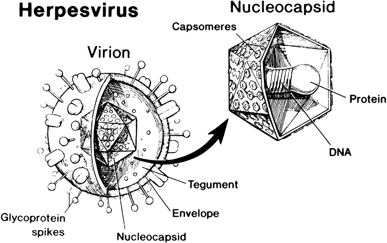
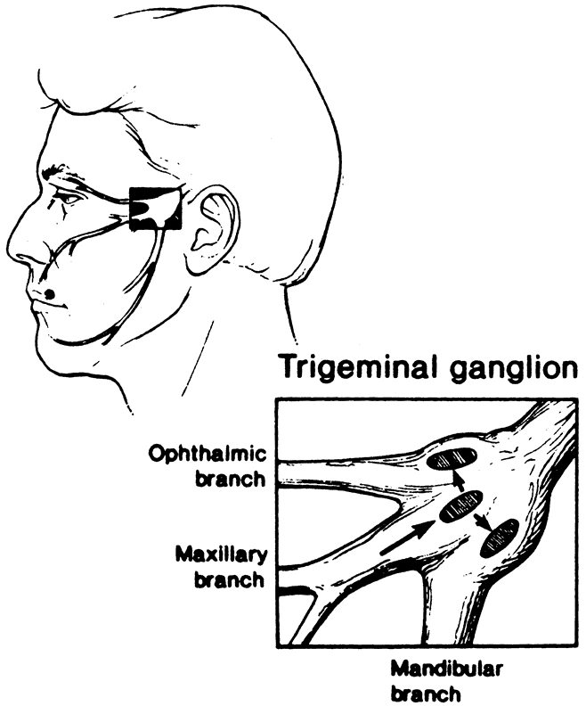
1. Liesegang TJ: Herpes simplex virus epidemiology and ocular importance. Cornea 20:1, 2001

2. Liesegang TJ, Melton LJ, Daly PJ et al: Epidemiology of ocular herpes simplex. Incidence in Rochester, MN, 1950 through 1982. Arch Ophthalmol 107:1155, 1989

3. Margolis TP, Atherton SS: Herpes simplex virus diseases: Posterior segment of the eye. In Pepose JS, Holland GN, Wilhelmus KR (eds): Ocular Infection and Immunity. St Louis: Mosby, 1996:1155

4. Roizman B: Herpesviridae. In Fields BN, Knipe DM, Howley PM, et al (eds): Fields Virology. Philadelphia: Lippincott-Raven, 1996:2221

5. Liesegang TJ: Biology and molecular aspects of herpes simplex and varicella-zoster virus infections. Ophthalmology 99:781, 1992

6. Roizman B, Sears AE: Herpes simplex viruses and their replication. In Fields BN, Knipe DM, Howley PM, et al (eds): Fields Virology. Philadelphia: Lippincott-Raven, 1996:2231

7. Spear PG: Glycoproteins specified by herpes simplex virus. In Roizman B (ed): The Herpesviruses. New York: Plenum, 1985:315

8. McGeoch DJ, Dolan A, Donald S et al: Sequence determination and genetic content of the short unique region in the genome of herpes simplex virus type 1. J Mol Biol 181:1, 1985

9. McGeoch DJ, Dalrymple MA, Davision AF et al: The complete DNA sequence of the long unique region in the genome of herpes simplex virus type 1. J Gen Virol 69:1531, 1988

10. Buchman TG, Simpson T, Nosal C et al: The structure of herpes simplex virus DNA and its application to molecular epidemiology. Ann N Y Acad Sci 354:279, 1980

11. Centifanto-Fitzgerald YM, Yamaguchi T, Kaufman HE et al: Ocular disease pattern induced by herpes simplex virus is genetically determined by a specific region of viral DNA. J Exp Med 155:475, 1982

12. Smeraglia R, Hochadel J, Varnell ED et al: The role of herpes simplex virus secreted glycoproteins in herpetic keratitis. Exp Eye Res 35:443, 1982

13. Tullo AB, Coupes D, Klapper P et al: Analysis of glycoproteins expressed by isolates of herpes simplex virus causing different forms of keratitis in man. Curr Eye Res 6:33, 1987

14. Dix RD, McKendall RR, Baringer JR: Comparative neurovirulence of herpes simplex virus type 1 strains after peripheral or intracerebral inoculation of BALB/c mice. Infect Immun 40:103, 1983

15. Thompson RL, Cook ML, Devi-Rao GB et al: Functional and molecular analysis of the avirulent wild-type herpes simplex virus type 1 strain KOS. J Virol 58:203, 1986

16. Thompson RL, Devi-Rao GV, Stevens JG et al: Rescue of a herpes simplex virus type 1 neurovirulence function with a cloned DNA fragment. J Virol 55:504, 1985

17. Thompson RL, Rogers SK, Zerhusen MA: Herpes simplex virus neurovirulence and productive infection of neural cells is associated with a function which maps between 0.82 and 0.832 map units on the HSV genome. Virology 172:435, 1989

18. Chou J, Kern E, Whitley RJ et al: Mapping of neurovirulence to protein 31.5 encoded by a diploid herpes simplex virus type 1 gene nonessential for growth in cell culture. Science 250:1262, 1990

19. Whitley RJ, Kern ER, Chatterjee S et al: Replication, establishment of latency, and induced reactivation of herpes simplex virus γ1 34.5 deletion mutants in rodent models. J Clin Invest 91:2837, 1993

20. Chou J, Chen JJ, Gross M et al: Association of Mr 90,000 phosphoprotein with protein kinase PKR in cells exhibiting enhanced phosphorylation of translation initiation factor eIF-2α and premature shutoff of protein synthesis after infection with γ134.5- mutants of herpes simplex virus 1. Proc Natl Acad Sci U S A 92:10516, 1995

21. Black FL: Infectious diseases in primitive societies. Science 187:515, 1975

22. Whitley RJ: Herpes simplex viruses. In Fields BN, Knipe DM, Howley PM, et al (eds): Fields Virology. Philadelphia: Lippincott-Raven, 1996:2297

23. Klein RJ: Pathogenetic mechanisms of recurrent herpes simplex virus infections. Arch Virol 51:1, 1976

24. Fenwick M, Morse LS, Roizman B: Anatomy of herpes simplex virus DNA. XI. Apparent clustering of functions effecting rapid inhibition of host DNA and protein synthesis. J Virol 29:825, 1979

25. Honess RW, Roizman B: Regulation of herpevirus macromolecular synthesis. Part I. Cascade regulation of synthesis of viral proteins. J Virol 14:8, 1974

26. McKnight JLC, Kristie TM, Roizman B: Binding of the virion protein mediating α gene induction in herpes simplex virus 1-infected cells to its cis site requires cellular proteins. Proc Natl Acad Sci U S A 84:7061, 1987

27. Jacob RJ, Roizman B: Anatomy of herpes simplex virus DNA. XIII. Accumulation of head to tail concatamers in nuclei of infected cells and their role in the generation of the four isomeric arrangement of viral DNA. J Virol 23:394, 1977

28. Katz JP, Boden ET, Coen DM: Quantitative polymerase chain reaction analysis of herpes simplex virus DNA in ganglia of mice infected with replication incompetent mutants. J Virol 64:4288, 1990

29. Wagner EK, Bloom DC: Experimental investigation of herpes simplex virus latency. Clin Microbiol Rev 10:419, 1997

30. Wagner EK, Bloom DC: HSV gene expression during latent infection and reactivation. In Medveczky PG, Friedman H, Bendinelli M (eds): Herpesviruses and Immunity. New York: Plenum, 1998:53

31. Ellerick DM, Fraser NW: Physical state of the latent herpes simplex virus genome in a mouse model system: Evidence suggesting an episomal state. Virology 158:265, 1987

32. Dix RD, Mills J: Experimental mouse models of herpes simplex virus infection. In Zak O, Sande MA (eds): Experimental Models in Antimicrobial Chemotherapy. Vol 2. London: Academic, 1986:219

33. Lillycrop KA, Dent CL, Wheatley S et al: The octamer-binding protein Oct-2 represses HSV immediate-early genes in cell lines derived from latently infectible sensory neuron. Neuron 7:381, 1991

34. Stevens JG, Wagner EK, Devi-Rao GB et al: RNA complementary to a herpesvirus α gene mRNA is prominent in latently 
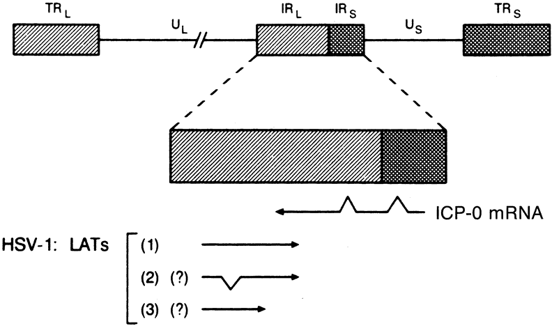
infected neurons. Science 235:1056, 1987

35. Stevens JG, Haarr L, Porter DD et al: Prominence of the herpes simplex virus latency-associated transcript in trigeminal ganglia from seropositive humans. J Infect Dis 158:117, 1988

36. Krause PR, Croen KD, Straus SE et al: Detection and preliminary characterization of herpes simplex virus type 1 transcripts in latently infected human trigeminal ganglia. J Virol 62:4819, 1988

37. Fraser NW, Block TM, Spivack JG: The latency-associated transcripts of herpes simplex virus: RNA in search of function. Virology 191:1, 1992

38. Farrel MJ, Dobson AT, Feldmen LT: Herpes simplex virus latency-associated transcript is a stable intron. Proc Natl Acad Sci U S A 88:790, 1991

39. Rock DL, Nesburn AB, Ghiasi H et al: Detection of latency related viral RNAs in trigeminal ganglia of rabbits latently infected with herpes simplex virus type 1. J Virol 61:3820, 1987

40. Perng G-C, Jones C, Ciacci-Zanella J et al: Virus-induced neuronal apoptosis blocked by the herpes simplex virus latency-associated transcript. Science 287:1500, 2000

41. Inman M, Perng G-C, Herderson G et al: Region of herpes simplex virus type 1 latency-associated transcript sufficient for wild-type spontaneous reactivation promotes cell survival in tissue culture. J Virol 75:3636, 2001

42. Thompson RL, Sawtell NM: Herpes simplex virus type 1 latency-associated transcript gene promotes neuronal survival. J Virol 75:6660, 2001

43. Ahmed M, Lock M, Miller CG et al: Regions of the herpes simplex virus type 1 latency-associated transcript that protect cells from apoptosis in vitro and protect neuronal cells in vivo. J Virol 76:717, 2002

44. Henderson S, Huen D, Rowe M et al: Epstein-Barr virus-coded BHRF1 protein, a viral homologue of Bcl-2, protects human B-cells from programmed cell death. Proc Natl Acad Sci U S A 90:8479, 1993

45. Derfuss T, Fickenscher H, Kraft MS et al: Antiapoptotic activity of the herpesvirus saimiri-encoded Bcl-2 homolog: Stabilization of mitochondria and inhibition of caspase 3-like activity. J Virol 72:5897, 1998

46. Aubert M, Blaho J: The herpes simplex virus type 1 regulatory protein ICP27 is required for the prevention of apoptosis in infected human cells. J Virol 73:2803, 1999

47. Ho DY, Mocarski ES: Herpes simplex virus latent RNA (LAT) is not required for latent infection in the mouse. Proc Natl Acad Sci U S A 86:7596, 1989

48. Sederati F, Izumi KM, Wagner EK et al: Herpes simplex virus type 1 latency associated transcription plays no role in establishment or maintenance of a latent infection in murine sensory neurons. J Virol 63:4455, 1989

49. Halford WP, Schaffer PA: ICP0 is required for efficient reactivation of herpes simplex virus type 1 from neuronal latency. J Virol 75:3240, 2001

50. Abghari SZ, Stulting RD: Recovery of herpes simplex virus from ocular tissues of latently infected inbred mice. Invest Ophthalmol Vis Sci 29:239, 1988

51. Kaye SM, Lynas C, Patterson A et al: Evidence for herpes simplex viral latency in the human cornea. Br J Ophthalmol 75:195, 1991

52. Easty DL: Herpes simplex virus isolation in chronic stromal keratitis, human and laboratory studies. Curr Eye Res 6:69, 1987

53. Laylock KA, Lee SF, Stulting RD et al: Herpes simplex virus type 1 transcription is not detectable in quiescent human stromal keratitis by in situ hybridization. Invest Ophthalmol Vis Sci 34:285, 1993

54. Cleator GM, Klapper PE, Dennett C et al: Corneal donor infection by herpes simplex virus: Herpes simplex virus DNA in donor corneas. Cornea 13:294, 1994

55. Neufeld MV, Steinemann TL, Merin LM et al: Identification of a herpes simplex virus-induced dendrite in an eye-bank donor cornea. Cornea 18:489, 1999

56. Redpath S, Angulo A, Gascoigne NRJ: Immune checkpoints in viral latency. Annu Rev Microbiol 55:531, 2001

57. McKendall RR, Klassen T, Baringer JR: Host defenses in herpes simplex infections of the nervous system: Effect of antibody on disease and viral spread. Infect Immun 23:305, 1979

58. Oakes JE, Lausch RN: Role of Fc fragments in antibody-mediated recovery from ocular and subcutaneous herpes simplex virus infections. Infect Immun 33:109, 1981

59. Dix RD, Pereira L, Baringer JR: Use of monoclonal antibody directed against herpes simplex virus glycoproteins to protect mice against acute virus-induced neurological disease. Infect Immun 34:192, 1981

60. Rector ZT, Lausch RN, Oakes JE: Use of monoclonal antibodies for analysis of antibody-dependent immunity to ocular herpes simplex virus type 1 infection. Infect Immun 38:168, 1982

61. Rector JR, Lausch RN, Oakes JE: Identification of infected cell-specific monoclonal antibodies and their role in host resistance to ocular herpes simplex virus type 1 infection. J Gen Virol 65:657, 1984

62. Dix RD: Glycoprotein gB of herpes simplex virus expresses type-common and type-specific antigenic determinants in vivo. J Med Virol 30:192, 1990

63. Dix RD, Mills J: Acute and latent herpes simplex virus neurological disease in mice immunized with purified virus-specific glycoproteins gB or gD. J Med Virol 17:9, 1985

64. Blyth WA, Harbour DA, Hill TJ: Pathogenesis of zosterform spread of herpes simplex virus in the mouse. J Gen Virol 65:1477, 1984

65. Schmid DS, Rouse BT: The role of T cell immunity in control of herpes simplex virus. Curr Top Microbiol Immunol 179:57, 1992.

66. Smith G: Virus strategies for evasion of the host response to infection. Trends Microbiol 2:81, 1994

67. York IA, Roop C, Andrews DW et al: A cytosolic herpes simplex virus protein inhibits antigen presentation to CD8+ T lymphocytes. Cell 77:525, 1994

68. Hill A, Jugovic P, York I et al: Herpes simplex virus turns off the TAP to evade host immunity. Nature 375:411, 1995

69. Jones TR, Hanson LK, Sun L et al: Multiple independent loci within the human cytomegalovirus unique short region down-regulate expression of major histocompatibility complex class I heavy chains. J Virol 69:4830, 1995

70. Moore K, Vieira P, Fiorentino D et al: Homology of cytokine synthesis inhibitory factor (Il-10) to the Epstein-Barr virus gene BCRF1. Science 248:1230, 1990

71. Sundmacher R: Clinical aspects of herpetic eye diseases. Curr Eye Res 6:183, 1987

72. Pepose JS, Leib DA, Stuart M et al: Herpes simplex virus diseases: Anterior segment of the eye. In Pepose JS, Holland GN, Wilhelmus KR (eds): Ocular Infection and Immunity. St Louis: Mosby, 1996:905

73. Hendricks RL: Immunopathogenesis of viral ocular infections. Chem Immunol 73:120, 1999

74. Thomas J. Rouse BT: Immunopathogenesis of herpetic ocular disease. Immunol Res 16:375, 1997

75. Stulting RD, Kindle JC, Nahmias AJ: Patterns of herpes simplex keratitis in inbred mice. Invest Ophthalmol Vis Sci 26:1360, 1985

76. Chen W, Tang Q, Hendricks RL: Ex vivo model of leukocytes migrate into herpes simplex virus-infected mouse corneas. J Leukoc Biol 60:167, 1996

77. Thomas J, Gangappa S, Kanangat S et al: On the essential involvement of neutrophils in the immunopathologic disease: Herpetic stromal keratitis. J Immunol 158:1383, 1997

78. Tumpey TM, Chen SH, Oakes JE et al: Neutrophil-mediated suppression of virus replication after herpes simplex virus type 1 infection of the murine cornea. J Virol 70:898, 1996

79. Hendricks RL, Tumpey TM, Finnegan A: IFN-gamma and IL-2 are protective in the skin but pathologic in the corneas of HSV-1-infected mice. J Immunol 149:3023, 1992

80. Zheng M, Deshpande S, Lee S et al: Contribution of vascular endothelial growth factor in the neovascularization process during the pathogenesis of herpetic stromal keratitis. J Virol 75:9828, 2001

81. Metcalf JF, Hamilton DS, Reichert RW: Herpetic keratitis in athymic (nude) mice. Infect Immun 26:1164, 1979

82. Brandt CR: Susceptibility of +/+, +/v, and v/v BALB/c mice to ocular herpes simplex virus infection. Ophthalmic Res 24:332, 1992

83. Mercadal C, Bouley D, DeStephano D et al: Herpetic stromal keratitis in the reconstituted SCID mouse model. J Virol 67:3404, 1993

84. Russel RG, Naisse MP, Larsen HS et al: Role of T lymphocytes in the pathogenesis of herpetic stromal keratitis. Invest Ophthalmol Vis Sci 25:938, 1984

85. Von Herranth MG, Oldstone MBA: Virus-induced autoimmune disease. Curr Opin Immunol 8:878, 1996

86. Oldstone MBA: Molecular mimicry and immune-mediated diseases. FASEB J 12:1255, 1998

87. Whitton LJ, Fujinami RS: Viruses as triggers of autoimmunity: Facts and fantasies. Curr Opin Microbiol 2:392, 1999

88. Sercarz EE, Lehmann PV, Ametani A et al: Dominance and crypticity of T cell antigenic determinants. Annu Rev Immunol 11:729, 1993

89. Zhou A, Granucci F, Yeh L et al: Molecular mimicry by herpes simplex virus type-1: Autoimmune disease after viral infection. Science 279:1344, 1998

90. Avery AC, Zhao ZS, Rodriguez A et al: Resistance to herpes stromal keratitis conferred by an IgG2a derived peptide. Nature 276:431, 1995

91. Opremcak EM, Wells PA, Thompson P et al: Immunogenetic influence of IgH-1 phenotype on experimental herpes simplex virus type-1 corneal infection. Invest Ophthalmol Vis Sci 29:749, 1988

92. Thomas J, Rouse BT: Immunopathology of herpetic stromal keratitis: Discordance in CD4+ T cell function between euthymic host and reconstituted SCID recipients. J Immunol 160:3965, 1998

93. Deshpande SP, Lee S, Zheng M et al: Herpes simplex virus-induced keratitis: Evaluation of the role of molecular mimicry in lesion pathogenesis. J Virol 75:3077, 2001

94. Ghiasi H, Slanina S, Nesburn AB et al: Characterization of baculovirus-expressed herpes simplex virus type 1 glycoprotein K. J Virol 68:2347, 1994

95. Ghiasi H, Kaiwar R, Nesburn AB et al: Immunoselection of recombinant baculoviruses expressing high levels of biologically active herpes simplex virus type 1 glycoprotein D. Arch Virol 121:163, 1991

96. Ghiasi H, Kaiwar R, Nesburn AB et al: Expression of glycoprotein B of herpes simplex virus type 1 in insect cells: Analysis of its biochemical and immunological properties. Virus Res 22:25, 1992

97. Ghiasi H, Kaiwar R, Nesburn AB et al: Baculovirus expressed glycoprotein G of herpes simplex virus type 1 partially protects vaccinated mice against lethal HSV-1 challenge. Virology 190:233, 1992

98. Ghiasi H, Kaiwar R, Nesburn AB et al: Expression of herpes simplex virus type 1 glycoprotein I (gI) in baculovirus: Preliminary biochemical characterization and protective studies. J Virol 66:2505, 1992

99. Ghiasi H, Kaiwar R, Nesburn AB et al: Baculovirus expressed glycoprotein H (gH) of herpes simplex virus type 1 (HSV-1) induces neutralizing antibody and delayed hypersensitivity responses but does not protect immunized mice against lethal HSV-1 challenge. J Gen Virol 73:719, 1992

100. Ghiasi H, Kaiwar R, Nesburn AB et al: Baculovirus expressed herpes simplex virus type 1 glycoprotein C protects mice from lethal HSV-1 infection. Antiviral Res 18:291, 1992

101. Ghiasi H, Kaiwar R, Nesburn AB et al: Baculovirus expressed glycoprotein E (gE) of herpes simplex virus type-1 (HSV-1) protects mice against lethal intraperitoneal and lethal ocular HSV-1 challenge. Virology 188:469, 1992

102. Ghiasi H, Cai S, Nesburn AB et al: Vaccination with herpes simplex virus type 1 glycoprotein K impairs clearance of virus from the trigeminal ganglia resulting in chronic infection. Virology 224:330, 1996

103. Ghiasi H, Cai S, Slanina S et al: Nonneutralizing antibody against the glycoprotein K of herpes simplex virus type-1 exacerbates herpes simplex virus type-1-induced corneal scarring in various virus-mouse strain combinations. Invest Ophthalmol Vis Sci 38:1213, 1997

104. Ghiasi H, Hofman FM, Cai S et al: Vaccination with different HSV-1 glycoproteins induces different patterns of ocular cytokine responses following HSV-1 challenge of vaccinated mice. Vaccine 17:2576, 1999

105. Ghiasi H, Perng G-C, Nesburn AB et al: Antibody-dependent enhancement of HSV-1 infection by anti-gK sera. Virus Res 68:137, 2000

106. Littaua R, Kurane I, Ennis FA: Human IgG Fc receptor II mediates antibody-mediated enhancement of dengue virus infection. J Immunol 144:3183, 1990

107. Halstead SB: In vivo enhancement of dengue virus infection in rhesus monkeys by passively transferred antibody. J Infect Dis 140:527, 1979

108. Culbertson WW, Dix RD: Varicella-zoster virus diseases: Posterior segment of the eye. In Pepose JS, Holland GN, Wilhelmus KR (eds): Ocular Infection and Immunity. St Louis: Mosby, 1996:1131

109. Lewis ML, Culbertson WW, Post JD et al: Herpes simplex virus type 1. A cause of acute retinal necrosis syndrome. Ophthalmology 96:875, 1989

110. Duker JS, Nielsen JC, Eagle RC Jr et al: Rapidly progressive acute retinal necrosis secondary to herpes simplex virus, type 1. Ophthalmology 97:1638, 1990

111. De La Paz MA, Young LHY: Acute retinal necrosis syndrome. Semin Ophthalmol 8:61, 1993

112. el Azazi, Samuelsson A, Linde A et al: Intrathecal antibody production against viruses of the herpesvirus family in acute retinal necrosis syndrome. Am J Ophthalmol 112:76, 1991

113. von Szily A: Experimental endogenous transmission of infection from bulbus to bulbus. Klin Monatsbl Augenheilkd 75:593, 1924

114. Whittum JA, McCulley JP, Niederkorn JY et al: Ocular disease induced in mice by anterior chamber inoculation of herpes simplex virus. Invest Ophthalmol Vis Sci 25:1065, 1984

115. Hamasaki DI, Dix RD, Atherton SS: Bilateral alterations of the ERG and retinal histology following unilateral HSV-1 inoculation. Invest Ophthalmol Vis Sci 29:1242, 1988

116. Atherton SS, Streilein SW: Two waves of virus following anterior chamber inoculation HSV-1. Invest Ophthalmol Vis Sci 28:571, 1987

117. Margolis TP, LaVail JH, Setzer PY et al: Selective spread of herpes simplex virus in the central nervous system after ocular infection. J Virol 63:4756, 1989

118. Vann VR, Atherton SS: Neural spread of herpes simplex virus after anterior chamber inoculation. Invest Ophthalmol Vis Sci 32:2462, 1991

119. Olson RM, Holland GN, Goss SJ et al: Routes of viral spread in the von Szily model of herpes simplex virus retinopathy. Curr Eye Res 6:59, 1987

120. Cousins SW, Gonzalez A, Atherton SS: Herpes simplex retinitis in the mouse: Clinicopathologic correlations. Invest Ophthalmol Vis Sci 30:1485, 1989

121. Azumi A, Cousins SW, Kanter MY et al: Modulation of murine herpes simplex virus type 1 retinitis in the uninoculated eye by CD4+ T lymphocytes. Invest Ophthalmol Vis Sci 35:54, 1994

122. Azumi A, Atherton SS: Sparing of the ipsilateral retina following anterior chamber inoculation of HSV-1: Requirement for either CD4+ or CD8+ T cells. Invest Ophthalmol Vis Sci 35:3251, 1994

123. Berra A, Rodriguez A, Heiligenhaus A et al: The role of macrophages in the pathogenesis of HSV-1 induced chorioretinitis in BALB/c mice. Invest Ophthalmol Vis Sci 35:2990, 1994

124. Streilein JW, Igietseme JU, Atherton SS: Evidence that precursor cytotoxic T cells mediate acute necrosis in HSV-1-infected retinas. Curr Eye Res 10:81, 1991

125. Zaltas MM, Opremcak M, Hemady R et al: Immunohistopathologic findings in herpes simplex virus chorioretinitis in the von Szily model. Invest Ophthalmol Vis Sci 33:68, 1992

126. Atherton SS, Altman NH, Streilein JW: Histopathologic study of herpes virus-induced retinitis in athymic BALB/c mice: Evidence for an immunopathologic process. Curr Eye Res 8:1179, 1989

127. Cousins SW, Altman NH, Atherton SS: Schisis contributes to necrosis in experimental HSV-1 retinitis. Exp Eye Res 48:745, 1987

128. Igietseme JU, Calzada PJ, Gonzalez AR et al: Protection of mice from herpes simplex virus-induced retinitis by in vitro-activated immune cells. J Virol 63:4808, 1989

129. Zhao M, Azumi A, Atherton SS: T lymphocyte infiltration in the brain following anterior chamber inoculation of HSV-1. J Neuroimmunol 58:11, 1995

130. Atherton SS: Protection from retinal necrosis by passive transfer of monoclonal antibody specific for herpes simplex virus glycoprotein D. Curr Eye Res 11:45, 1992

131. Zhao M, Atherton SS: Immune effector cell (IEC)-mediated protection from HSV-1 retinitis occurs in the brain. J Neuroimmunol 75:51, 1997

132. Dix RD, Hamasaki DI, Hurst L et al: Bilateral retinal disease following unilateral inoculation of mice with a HSV-1 isolate from a patient with acute retinal necrosis. Invest Ophthalmol Vis Sci 31:314, 1990

133. Streilein JW: Regional immunity of the eye. In Pepose JS, Holland GN, Wilhelmus KR (eds): Ocular Infection and Immunity. St Louis: Mosby, 1996:19

134. Niederkorn JY, Ferguson TA: Anterior chamber associated immune deviation (ACAID). In Pepose JS, Holland GN, Wilhelmus KR (eds): Ocular Infection and Immunity. St Louis: Mosby, 1996:96

135. Streilein JW, Atherton SS, Vann V: A critical role for ACAID in the distinctive pattern of retinitis that follows anterior chamber inoculation of HSV-1. Curr Eye Res 6:127, 1987

136. Kielty D, Cousins SW, Atherton SS: HSV-1 retinitis and delayed hypersensitivity in DBA/2 and C57BL/6 mice. Invest Ophthalmol Vis Sci 28:1994, 1987

137. Chodosh J, Banks MC, Stroop WG: Rose bengal inhibits herpes simplex virus replication in Vero and human corneal epithelial cells in vitro. Invest Ophthalmol Vis Sci 33:2520, 1992

138. Brooks SE, Kaza V, Nakamura T et al: Photo inactivation of herpes simplex virus by rose bengal and fluorescein. In vitro and in vivo studies. Cornea 13:43, 1994

139. Chodosh J, Dix RD, Howell RC et al: Staining characteristics and antiviral activity of sulforhodamine B and lissamine green B. Invest Ophthalmol Vis Sci 35:1046, 1994

140. Stroop WG, Chen MC, Chodosh J et al: Polymerase chain reaction assessment of herpes simplex virus type 1 corneal infection in animals treated with rose bengal and lissamine green B. Invest Ophthalmol Vis Sci 41:2096, 2000

141. Waldrep JC, Mondino BJ: Humoral immunity and the eye. In Pepose JS, Holland GN, Wilhelmus KR (eds): Ocular Infection and Immunity. St Louis: Mosby, 1996:33

142. Goldmann H, Witmer R: Antikurper in Kammerwasser. Ophthalmologica 177:323, 1954

143. Witmer R: Clinical implications of aqueous humor studies in uveitis. Am J Ophthalmol 86:39, 1978

144. Mullis K, Faloona F, Scharf S et al: Specific enzymatic amplification of DNA in vitro: The polymerase chain reaction. Symp Quant Biol 51:263, 1986

145. Fox GM, Crouse CA, Chuang EL et al: Detection of herpevirus DNA in vitreous and aqueous specimens by the polymerase chain reaction. Arch Ophthalmol 109:266, 1991

146. Grant DM: Acyclovir (Zovirax) ophthalmic ointment: A review of clinical tolerance. Curr Eye Res 6:231, 1987

147. Charles SJ, Gray JJ: Ocular herpes simplex virus infection: Reduced sensitivity to acyclovir in primary disease. Br J Ophthalmol 74:286, 1990

148. Sonkin PL, Baratz KH, Frothingham R et al: Acyclovir-resistant herpes simplex virus keratouveitis after penetrating keratoplasty. Ophthalmology 99:1805, 1992

149. Dix RD: Prospects for a vaccine against herpes simplex virus types 1 and 2. Prog Med Virol 34:89, 1987

150. Arvin AM: Varicella-zoster virus. In Fields BN, Knipe DM, Howley PM, et al (eds): Fields Virology. Philadelphia: Lippincott-Raven, 1996:2547

151. Hammerschlag MR, Gershon AA, Steinberg SP et al: Herpes zoster in an adult recipient of live attenuated varicella vaccine. J Infect Dis 160:535, 1989

152. Plotkin SA, Starr SE, Connor K et al: Zoster in normal children after varicella vaccine. J Infect Dis 159:1000, 1989

153. Gershon AA: Live attenuated varicella vaccine use in immunocompromised children and adults. Pediatrics 78:757, 1986

154. Plummer G: Serological comparison of the herpes viruses. Br J Exp Pathol 45:135, 1964

155. Mitchell WJ, Deshmane SL, Dolan A et al: Characterization of herpes simplex virus type 2 transcription during latent infection of mouse trigeminal ganglia. J Virol 64:5342, 1990

156. Neumann-Haefelin D, Sundmacher R, Wochnik G et al: Herpes simplex virus types 1 and 2 in ocular disease. Arch Ophthalmol 96:64, 1978

157. Rosenwasser GOD, Greene WH: Simultaneous herpes simplex virus type 1 and 2 keratitis in acquired immunodeficiency syndrome. Am J Ophthalmol 113:102, 1992

158. Ganatra JB, Chandler D, Santos C et al: Viral causes of the acute retinal necrosis syndrome. Am J Ophthalmol 129:166, 2000

159. Van Gelder RN, Willig JL, Holland GN et al: Herpes simplex virus type 2 as a cause of acute retinal necrosis syndrome in young patients. Ophthalmology 108:869, 2001

160. Itoh N, Matsumura N, Ogi A et al: High prevalence of herpes simplex virus type 2 in acute retinal necrosis syndrome associated with herpes simplex virus in Japan. Am J Ophthalmol 129:404, 2000

Back to Top