Chapter 113D The Role of Fluorescein Angiography in Diabetic Retinopathy R. JOSEPH OLK and CAROL M. LEE Table Of Contents |
BACKGROUND DIABETIC RETINOPATHY UNEXPLAINED VISUAL LOSS MACULAR EDEMA PROLIFERATIVE DIABETIC RETINOPATHY PSEUDOPHAKIC VERSUS DIABETIC MACULAR EDEMA SUMMARY REFERENCES |
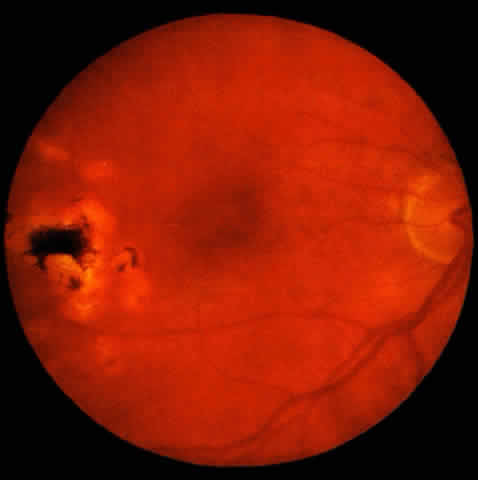
Diabetic retinopathy is the leading cause of blindness in Americans between 20 and 74 years of age.1 Fluorescein angiography has helped in the diagnosis, management, and study of the pathophysiologic mechanisms of this disease. The specific indications for fluorescein angiography will be described in this chapter, underscoring the established basic guidelines outlined in the recently published Preferred Practice Pattern by the American Academy of Ophthalmology.1 |
BACKGROUND DIABETIC RETINOPATHY |
Fluorescein angiography is not indicated as a screening tool for diabetic
retinopathy nor as a baseline measurement of background diabetic retinopathy. The
signs of background diabetic retinopathy include microaneurysms, intraretinal
hemorrhages, hard exudates, and macular edema, all
of which should be visible on clinical examination.2–8 Ophthalmoscopy of a well-dilated fundus performed by trained ophthalmologists
and technical personnel has been shown to agree with fundus photography
in at least 85% of cases; disagreement occurred in cases of
the earliest stages of retinopathy whereby a photographically observable
microaneurysm may not be visualized ophthalmoscopically.9,10 The risk of underevaluating retinopathy consistently occurred in eyes
with minimal changes. Clinical identification of more severe changes was
almost always consistent and in agreement with the photographic findings. Stereo fundus photography provides a permanent record of the fundus appearance and can be used to document the severity and progression of disease.1,5,6,9–11 The seven standard fields of stereo color fundus photography as specified by the modified Airlie House classification3 allows for both detailed grading and documentation of progression of disease, or conversely, a response to treatment. Fundus photography is clearly valuable in documenting retinopathy that is changing rapidly, or immediately prior to treatment, once the decision to treat has been made on clinical grounds. Large 60° fields will give a more complete view of the midperipheral regions than 30° photography and may be the field of choice for scanning for retinopathy. If fundus photography is considered to be 100% sensitive in detecting retinopathy, ophthalmoscopy correlates with retinopathy detected by photography 86% of the time.9 For proliferative diabetic retinopathy, between 79% to 96% agreement was seen between ophthalmoscopic examination and fundus photography.9,10 This assumes, however, a dilated fundus examination, as the sensitivity of ophthalmoscopy has been shown to diminish by 50% in an undilated eye.12 Given the high specificity and sensitivity of the clinical ophthalmoscopic examination, several screening strategies have been studied.5,6 An annual dilated ophthalmoscopic examination for eyes with no retinopathy has been recommended in several cost-effectiveness models.1,5,6 Although in such a cost-effectiveness model photography does not enhance the outcome, fundus photographs are nevertheless medically useful for documenting the course of disease. |
UNEXPLAINED VISUAL LOSS | ||
Fluorescein angiography can be helpful in determining the cause of unexplained
visual loss in an eye with visual complaints out of proportion
to the retinopathy clinically seen.1,8,13,14 An eye may have both minimal peripheral changes and minimal macular edema
but visual acuity far worse than the clinical examination. Causes
may include an optic neuropathy or a markedly ischemic macula, the latter
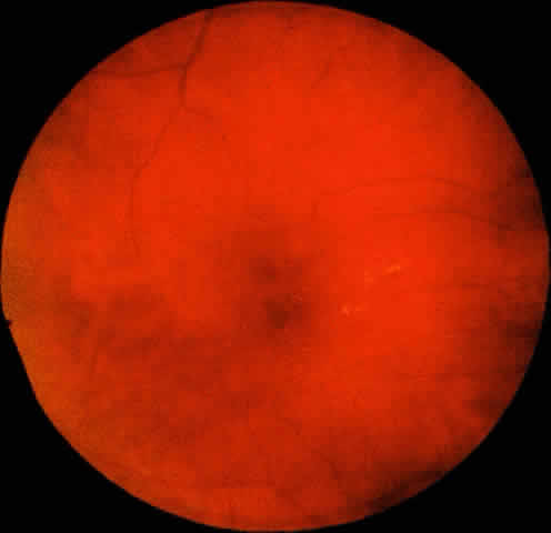
of which could be confirmed by angiography (Color Plate. 1, Fig. 1).
Occlusion of the perifoveal capillaries is readily shown on angiography2,4,13,14 as abnormalities of the foveal avascular zone (FAZ). These angiographic characteristics include an irregularity of the margins of the FAZ, budding of capillaries into the FAZ, and the development of wide intercapillary spaces within bridging vessels in the perifoveal capillary bed.2,13 The perifoveal capillaries may actually be more apparent due to both dilation of the remaining capillaries and their contrast to the larger zone of nonperfusion seen angiographically as hypofluorescence. The enlargement of the FAZ is presumed to cause a loss of visual acuity due to macular ischemia.1,2,13–15 |
MACULAR EDEMA | ||||||
Macular edema is the leading cause of visual loss in eyes with diabetic
retinopathy.15 Numerous randomized clinical trials have demonstrated the efficacy of
laser photocoagulation in preventing visual loss due to diabetic macular
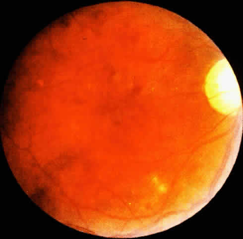
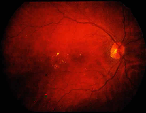
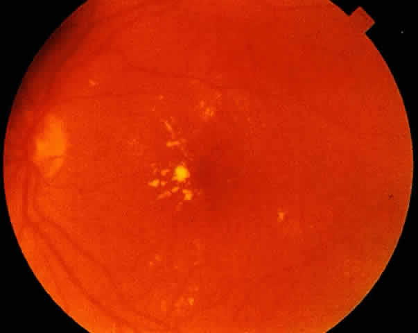
edema.15–21 The Early Treatment Diabetic Retinopathy Study (ETDRS)19,22–24 stated that patients with clinically significant macular edema be considered for treatment. They have shown that focal treatment substantially reduces the risk of visual loss from clinically significant diabetic macular edema regardless of initial visual acuity. Any of the following characteristics fulfills the definition of clinical significance: thickening of the retina at or within 500 μm of the center of the macula; hard exudates at or within 500 μm of the center of the macula, if associated with thickening of adjacent retina (not residual hard exudates remaining after disappearance of retinal thickening); or a zone or zones of retinal thickening one disc area or larger, any part of which is within one disc diameter of the center of the macula. The diagnosis of clinically significant macular edema is based on examination by stereo slit-lamp biomicroscopy and/or stereo fundus photography. Fluorescein angiography is not indicated to diagnose the presence of clinically significant macular edema. Angiographic evidence of fluorescein leakage without clinical evidence of retinal thickening does not fulfill the criteria for clinical significance. However, once the diagnosis and the decision to treat macular edema have been made, fluorescein angiography is extremely useful in helping to guide the treatment pattern. Angiography will identify treatable lesions and delineate the foveal avascular zone and the status of the macular perfusion. Fluorescein angiography will also confirm the presence of thickening and edema by leakage in the late stage of the angiogram, which also can be used as a guide to treatment (Color Plate. 2 ,Fig. 2).
Although the ETDRS showed a treatment benefit at all levels of visual acuity, if an eye has good vision and is asymptomatic, one can elect to follow the patient, rather than treat immediately. That patient should be followed at close intervals at least every 3 months; a fluorescein angiogram is not indicated until the decision to treat has been made based on the clinical examination (Color Plate. 3, Fig. 3).
The ETDRS has emphasized the use of focal treatment in the treatment of diabetic macular edema characterized by focal leakage. Diffuse diabetic macular edema may present a different pathophysiologic mechanism. Focal edema is a product mainly of individual leaking microaneurysms, whereas diffuse diabetic macular edema may be produced by a combination of a breakdown of the inner blood retinal barrier at the level of the retinal capillaries and arterioles and at the outer blood retinal barrier at the level of the retinal pigment epithelium.2,25 Other treatment protocols including Olk's modified grid photocoagulation emphasize the use of grid treatment for diffuse macular edema greater than 2 disc diameters in area and involving any portion of the foveal avascular zone.20,21,26 Again the diagnosis of diffuse macular edema is made on the clinical biomicroscopic examination, whereas the fluorescein angiogram is obtained only after the decision to treat has been made, and is used as a guide to treatment (Color Plate. 4, Fig. 4).
Supplemental macular treatment was required by the ETDRS if macular edema involved or threatened the center of the macula. Supplemental treatment was allowed for any degree of edema, which met one of the definitions for clinical significance.19,22,23 Fluorescein angiography is again useful in guiding the treatment once the clinical need for retreatment has been determined. Supplemental modified grid macular photocoagulation is recommended by Olk if residual central thickening involving the foveal avascular zone is seen on clinical examination.20,21,26 Only if retreatment is being considered does one obtain a fluorescein angiogram to delineate the areas of leakage, to identify the foveal avascular zone, and to guide in the pattern of treatment (Color Plate. 4, Fig. 4). |
PROLIFERATIVE DIABETIC RETINOPATHY | ||
The Diabetic Retinopathy Study (DRS) has determined four retinopathy risk
factors: the presence of new vessels, the location of new vessels on
or within one disc diameter of the optic disc, the severity of new vessels, and
the presence of preretinal or vitreous hemorrhage.27–30 If an eye has both neovascularization of the disc (NVD) and neovascularization
elsewhere (NVE), the severity applies to the NVD because in eyes
with NVD, the presence of moderate to severe NVE did not further increase
the risk of severe visual loss. The presence of three or four
retinopathy risk factors characterizes an eye as being “high-risk”; these
characteristics include any of the following: NVD greater
than one fourth to one third disc diameter in area and vitreous or
preretinal hemorrhage associated with less extensive NVD or with NVE
greater or equal to one half disc diameter in area. The DRS has shown
the clear benefit of scatter panretinal photocoagulation to these high-risk
eyes in preventing severe visual loss from the consequences of proliferative
disease. Fluorescein angiography is not needed to diagnose
the presence of high-risk retinopathy. The clinical examination is highly
sensitive in detecting this stage of retinopathy and is adequate
for the determination of the need for photocoagulation.1,9–11 Color fundus photography, as previously stated, employing the seven standard
stereo fields may be helpful in gauging the response to therapy
once panretinal photocoagulation has been instituted. As it has been shown that diabetic macular edema may be exacerbated by panretinal photocoagulation,31–33 any eye that shows clinical evidence of retinal thickening falling within the definition of clinically significant macular edema should have a fluorescein angiogram to guide in the treatment of the macular edema either prior to or in combination with the panretinal treatment of the proliferative disease (Color Plate. 5, Fig. 5).
Fluorescein angiography may be helpful in searching for subtle patches of neovascularization or capillary nonperfusion when either significant preproliferative signs are present or proliferative disease is suspected and media opacity precludes a good view. In this last circumstance, fluorescein angioscopy may be more helpful in scanning the retina to detect areas of leakage from neovascularization; however, fluorescein angioscopy is not helpful in the confirmation nor the guidance of treatment for macular edema. Angiography may also be helpful in detecting specific fronds of neovascularization when blood from a vitreous hemorrhage obscures the view; this can guide the ophthalmologist to areas of active neovascularization that may be treated with focal ablation. In general, indications for retreatment include increasing neovascularization, new areas of neovascularization, new vitreous hemorrhage, and failure of the initial neovascularization to regress.1,30,34 Each of these findings should be determined on clinical examination and do not usually need fluorescein angiography for confirmation. However, the progression or regression of neovascularization can be readily followed by serial clinical examinations and on color fundus photography, and can be used as a permanent record for comparison. The significance of capillary nonperfusion in the midperipheral retina has been illustrated in the fluorescein angiographic studies by Shimizu.35,36 Clinically the angiographic demonstration of a significant amount of capillary nonperfusion or dropout may be helpful in those special circumstances where photocoagulation treatment is being considered for severe preproliferative or early proliferative disease. These special circumstances include accelerated retinopathy during pregnancy, imminent cataract surgery or worsening cataract, unreliable or inadequate follow-up, concomitant presence of carotid occlusive disease, and the comparative status of the fellow eye. |
PSEUDOPHAKIC VERSUS DIABETIC MACULAR EDEMA | ||
Fluorescein angiography may be helpful in differentiating between macular
edema secondary to pseudophakia or to diabetes, as the clinical diagnosis
between the two is often difficult. Typically, in pseudophakic
macular edema, cysts distributed in a petalloid pattern can be appreciated
by slitlamp biomicroscopy.37 Fluorescein angiography will usually show early dye leakage from the parafoveal
capillaries in a uniform fashion with the later phases revealing
a polycystic pattern of dye accumulation caused by serous exudation
into the extracellular space. In addition, the capillaries of the optic
nerve head will frequently leak dye in the later frames of the angiogram. Diabetic
macular edema, however, is sometimes accompanied by
a cystoid component such that the differential between the two can be
difficult (Color Plate. 6, Fig. 6).
|
SUMMARY |
In conclusion, fluorescein angiography is a useful guide in the treatment of clinically significant macular edema, in the means of evaluating an eye with unexplained visual loss, and sometimes in identifying subtle areas of neovascularization or capillary nonperfusion. It is not indicated for the diagnosis of either clinically significant diabetic macular edema or proliferative diabetic retinopathy. Nor is it used as a screening tool or a baseline examination in patients with diabetic retinopathy. |