Chapter 113e
Fluorescein and Indocyanine Green Angiography in Infectious and Inflammatory Diseases of the Retina and Choroid
STEVEN R. SANISLO, MARK S. BLUMENKRANZ and THIERRY C. VERSTRAETEN
Main Menu   Table Of Contents

Search

PRINCIPLES OF FLUORESCEIN
PRINCIPLES OF INDOCYANINE GREEN (ICG)
METHOD FOR ASSESSING MECHANISM OF VISUAL LOSS AND RESPONSE TO TREATMENT
QUANTITATION OF DISEASE SEVERITY
INFLAMMATORY RETINAL PIGMENT
CONCLUSIONS
REFERENCES

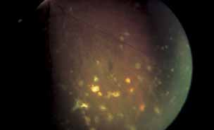
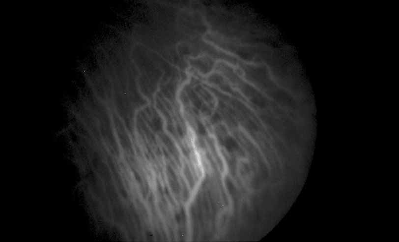
In many instances, angiography can serve as a useful guide to the clinician in the diagnosis and management of infectious and inflammatory diseases of the retina and choroid. Sodium fluorescein is the most common angiographic dye used for this purpose, but indocyanine green (ICG) dye is also useful in certain cases. Because angiography yields a high-contrast image for the permanent medical record, the potential exists for overuse of this modality, when more simple and inexpensive methods will suffice. In instances where clinical history and ophthalmoscopy alone are not sufficient to either establish a working diagnosis or develop and monitor a treatment plan, fluorescein or ICG angiography in conjunction with other serologic studies may play an important role. Because of restrictions on the length of this chapter, as well as the availability of excellent reviews on specific diseases, both in other portions of this series as well as elsewhere,1–4 an encyclopedic listing of the angiographic features of all major inflammatory and infectious conditions is not provided. Our intent is to establish basic guidelines for the indications and interpretation of fluorescein and ICG angiography in this broad category of diseases.
Back to Top
PRINCIPLES OF FLUORESCEIN

ANGIOGRAPHY

The rationale for the use of sodium fluorescein for diagnostic imaging of the fundus is covered more completely in other chapters. By absorbing photic energy in the wavelength range of 465 to 490 nm and emitting at a different wavelength range of 520 to 530 nm, sodium fluorescein yields a dynamic high-contrast image of the retinal structures, emphasizing the discrete intravascular compartments silhouetted against their adjacent structures, when photographed with appropriate barrier and excitation filters in place. In some instances, the presence of the monochromatic filters alone is sufficient to decrease light scattering and consequently improve resolution of fine detail in eyes with hazy intraocular media. In most instances, however, the luminescence provided by the excited fluorescein molecule is responsible for the apparent image enhancement available by this technique. With the recent availability of more sophisticated video and digital signal processing techniques, the capacity for image enhancement has been further increased. Because of its relatively low molecular weight of 376 Daltons, sodium fluorescein freely traverses the fenestrated capillaries and larger vessels comprising the choroid. However, the tight junctions between adjacent retinal pigment epithelial cells and healthy mature retinal capillary endothelial cells effectively block the ingress of fluorescein from the choroid to the subretinal space, or from the retinal vessels into the neurosensory retina or vitreous, respectively, under normal conditions. The loss of this selective barrier effect as a result of disease underlies the value of fluorescein angiography as an imaging modality. In addition to ischemia, mechanical or thermal trauma and inflammation result in breakdown of the barrier effect of tight junctions and allow passage of fluorescein, generally in proportion to the degree of the inflammation.1–4 Because the inflammation may be either generalized or localized to a specific region of the chorioretinal complex, depending on the nature of the disease process, the fluorescein angiogram may yield valuable information not only with regard to severity of the breakdown of the barrier such as fluorophotometry but also with regard to localization of the site of inflammation. In instances in which inflammation coexists with ischemia or neovascularization, fluorescein angiography can be particularly valuable in distinguishing between these elements. Additionally, fluorescein angiography can show alterations to the retinal pigment epithelium during acute inflammatory phases and also show chronic changes in the pigment epithelium that often remain after the inflammation has subsided.

Back to Top
PRINCIPLES OF INDOCYANINE GREEN (ICG)

ANGIOGRAPHY

Indocyanine green angiography differs from fluorescein angiography in several ways. Indocyanine green dye is more highly bound to protein than sodium fluorescein and has peak absorption and emission wavelengths in the infrared spectrum, outside of the visible range. Because the fluorescence efficiency of the ICG molecule is only 4% of that of sodium fluorescein, photographic film is too insensitive to record this weak fluorescence. To detect ICG fluorescence, charge coupled device (CCD) cameras with a digital acquisition process are necessary. Indocyanine green angiography can image the choroidal vasculature better than fluorescein angiography because the larger and more highly protein bound ICG molecule is unable to diffuse out of the fenestrated choroidal vessels. In addition, the infrared emission spectrum of ICG dye is not blocked by the overlying retinal pigment epithelium. Thus ICG angiography is an excellent technique for evaluating choroidal hypoperfusion or choroidal leakage.

Indocyanine green angiography can be helpful in the diagnosis and management of various infectious and inflammatory diseases. ICG patterns of hypofluorescence and hyperfluorescence have been described for many inflammatory diseases including: multiple evanescent white dot syndrome (MEWDS), 5–7 acute multifocal placoid pigment epitheliopathy (AMPPE),8 multi-focal choroiditis,9 Vogt-Koyanagi-Harada disease (VKH),10–12 ocular sarcoidosis,13 and Behcet disease.14,15 ICG angiography often shows discreet or patchy areas of hypofluorescence in the active inflammatory phases of these diseases indicative of choroidal alterations, often much more extensive than is clinically apparent with ophthalmoscopy or fluorescein angiography. In some of these disorders such as MEWDS the ICG, abnormalities resolve completely with resolution of the patient's symptoms, whereas in others ICG abnormalities may persist. Hyperfluorescence may also be seen with ICG angiography, as in VKH disease where diffuse choroidal leakage can be prominent in late phases of the study. By better-visualizing the condition of the choroidal circulation, ICG angiography is helping us to better-understand the pathophysiology and possible mechanisms involved in infectious and inflammatory diseases of the retina and choroid. Currently, the clinical usefulness of ICG angiography for infectious and inflammatory diseases remains somewhat limited. However, with increasing familiarity of the angiographic findings in these diseases, ICG angiography is likely to play a greater role in their diagnosis and management.

Back to Top
METHOD FOR ASSESSING MECHANISM OF VISUAL LOSS AND RESPONSE TO TREATMENT
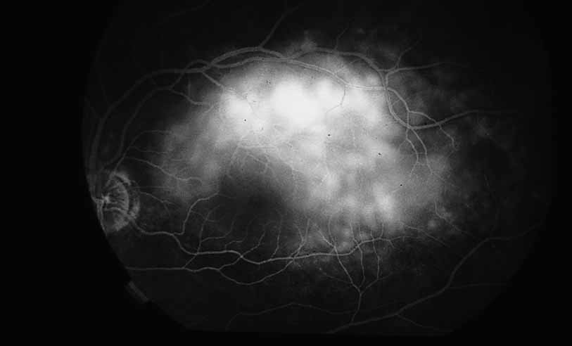
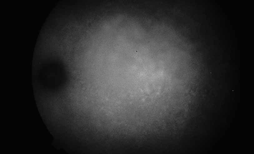
As indicated earlier, in the large majority of cases, the combination of the clinical history and ocular examination including binocular slit lamp biomicroscopy are generally sufficient to establish a working diagnosis and formulate a treatment plan. In selected instances, including the peculiar leopard spot appearance of luetic neuroretinitis16–21 or the classic brush-fire appearance of serpiginous choroiditis (see Figs. 1, 2, and 3),22–30 the diagnosis may be established on the basis of the angiographic findings alone. More often, the angiography is used to answer specific questions about the individual case, relating either to the mechanism of visual loss or to the measurement of the response to treatment. The following is a partial list of complications of inflammations and infections of the posterior segment that result in visual loss that can be better defined by angiography:

  Optic neuropathy
  Cystoid macular edema
  Retinal vascular occlusion
  Retinal (macular) capillary nonperfusion
  Retinal pigment epithelial detachment
  Retinal pigment epithelial dysfunction
  Retinal neurosensory detachment
  Choroidal neovascularization
  Choroidal vascular occlusion
  Neovascular glaucoma
  Vitreous hemorrhage

Fig. 1 Serpiginous choroiditis. Early frame of the fluorescein angiogram shows hypofluorescent and hyperflourescent patches extending outward from the optic nerve in a serpiginous pattern (Courtesy of Joseph Michaelson).

Fig. 2 Serpiginous choroiditis. Late frame of the fluorescein angiogram shows extensive staining of previously hypofluorescent zones, with continued hypofluorescence, characteristic of the acute phases of serpiginous choroiditis (Courtesy of Joseph Michaelson).

Fig. 3 Luetic neuroretinitis. Early-frame angiogram of eye of middle-aged man who presented with vision loss and a peculiar yellow---green discoloration of the outer retina and pigment epithelium. The patient was not known to be immunosuppressed, and subsequent serologic testing confirmed the presence of a positive VDRL and FTA-ABS. The early frame of the angiogram demonstrates a broad area of hypofluorescence corresponding to the clinical lesion. Note the subtle punctate leopard spots above the lesion.

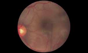
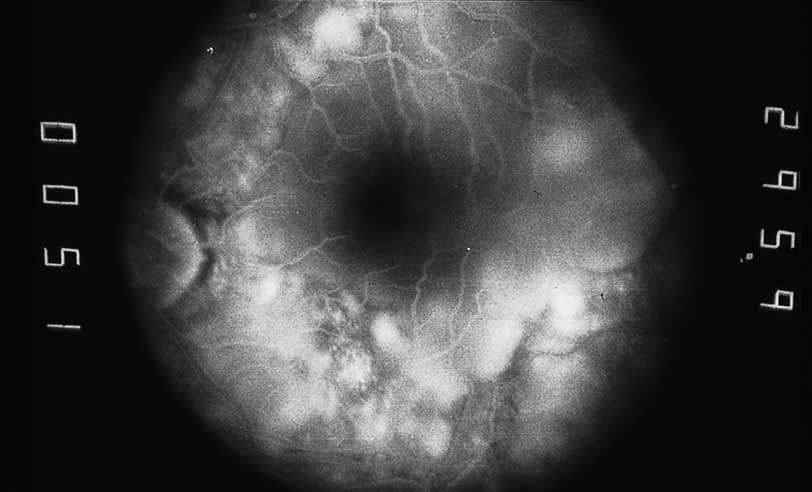
As an example, patients with posterior segment involvement by the spirochete Treponema pallidum may manifest visual loss on the basis of a large number of different mechanisms.16–21 These include perioptic neuritis, retinal periphlebitis or periarteritis, diffuse neuroretinitis, cystoid macular edema, or choroidal neovascularization. Fluorescein angiography may be helpful not only in establishing the causative agent but also in determining the predominant mechanism of visual loss (see Figs. 4 and 5).

Fig. 4 Luetic neuroretinitis. Color photograph showing placoid opacity at the level of the retinal pigment epithelium.

Fig. 5 Luetic neuroretinitis. A late frame of the angiogram in Figure 4 shows extensive staining of the entire area corresponding to leakage into the outer retina and staining of the pigment epithelium. The patient was successfully treated with intravenous penicillin therapy and subsequently developed diffuse “leopard spot” changes in the pigment epithelium characteristic of resolved luetic neuroretinitis.

Back to Top
QUANTITATION OF DISEASE SEVERITY

MACULAR INVOLVEMENT

One of the most useful applications of fluorescein angiography in infections and inflammations of the posterior segment is the delineation and quantization of cystoid macular edema. Although it is generally possible to ascertain the presence or absence of gross cystoid macular edema in eyes with clear media, this is often not the case in eyes with intraocular inflammation. The presence of intraocular inflammation, such as the vitreous cells accompanying pars planitis31–33 or sarcoidosis, 34–40 may prevent clear viewing of the macula. The tell-tale hyperfluorescence seen in the normally hypofluorescent macula after dye injection is an important indicator. The presence of cystoid macular edema by angiography may lead the clinician to take a more aggressive posture in the treatment of these diseases, especially in younger patients in whom some uncertainty exists regarding the relative contribution of the media opacity versus the macular function to the reduced vision (see Figs. 6 and 7).

Fig. 6 Presumed ocular histoplasmosis. This patient developed decreased vision with mild vitritis. Color photograph shows a hazy view of the central macula.

Fig. 7 Presumed ocular histoplasmosis. Fluorescein angiogram of Figure 6 demonstrates cystoid macular edema as the cause of this patient's decreased vision.

Once a treatment regimen has been instituted, the follow-up fluorescein angiogram can serve as a marker for treatment response in addition to other clinical variables, including visual acuity and severity of intraocular inflammation (see Fig. 8).

Fig. 8 Presumed ocular histoplasmosis. Repeat fluorescein angiogram from Figure 7 shows complete resolution of cystoid macular edema after intravitreal injection of triamcinolone–acetamide.

OPTIC NERVE DISEASE

The fluorescein angiogram can provide useful information with regard to inflammatory involvement of the optic nerve in association with retinal inflammation either in eyes with clear or opaque media. In eyes with clear media, excessive staining of the optic nerve in the late frames of the angiogram may signify sub clinical inflammation, even in the absence of any obvious disc swelling or engorgement. Patients with Vogt-Koyanagi-Harada disease, 41 sympathetic ophthalmia42–46 and luetic neuroretinitis frequently have occult inflammation of the optic nerve, which may signify the clinical stage of the intraocular inflammation more accurately than the retinal findings, particularly in long-standing cases with extensive atrophic retinal abnormalities in which it is difficult to monitor change (see Fig. 9).

Fig. 9 Luetic neuroretinitis. Color photographs show diffuse “leopard spot” scarring of the retinal pigment epithelium from previous retinitis. Fluorescein angiography shows persistent staining of the optic nerve (Courtesy of Joseph Michaelson).

Another circumstance in which fluorescein angiography is helpful in defining the cause and extent of inflammatory disease is in the context of a patient who has a coexisting disease process. Patients with diabetes mellitus who are found to have cystoid macular edema after cataract extraction represent a difficult management problem. Although diffuse leakage may occur from the perifoveal capillary net, in response to hyperglycemia or low-grade intraocular inflammation such as that which follows cataract surgery, the presence of late staining of the optic nerve in a fluorescein angiogram in such a patient suggests that at least a portion of the edema may be related to postsurgical inflammation.

A third instance in which fluorescein angiography concentrating on the optic nerve may be helpful is in eyes with vascular engorgement of the optic nerve, especially with media opacities. Fluorescein angiography may help the clinician distinguish abnormally leaking capillaries from neovascularization. In acute multifocal hemorrhagic retinal vasculitis, optic nerve involvement is common in the early stages of the disease, 47 with optic nerve disc neovascularization seen later (see Figs. 10, 11, 12, and 13). Another fluorescein angiographic clue to the distinction between optic disc vessel engorgement and true neovascularization is the presence of associated large zones of retinal capillary nonperfusion such as those seen in sarcoidosis (see Figs. 14 and 15),34–40 acute multifocal hemorrhagic retinal vasculitis (see Figs. 10 and 11),47 or Eale disease (see Figs. 16 and 17).48–52

Fig. 10 Acute multifocal hemorrhagic retinal vasculitis. A middle-aged man presented with the picture of multiple branch vein obstructions and low-grade intraocular inflammation.

Fig. 11 Acute multifocal hemorrhagic retinal vasculitis. Fluorescein angiogram confirms the presence of associated retinal capillary non-perfusion without neovascularization at the onset of the disease.

Fig. 12 Acute multifocal hemorrhagic retinal vasculitis. The patient later developed disk neovascularization with vitreous hemorrhage. Note the areas of neovascular leakage from the optic nerve.

Fig. 13 Acute multifocal hemorrhagic retinal vasculitis. The patient underwent scatter laser photocoagulation to the zones of retinal capillary nonperfusion, which resulted in regression of the neovascularization.

Fig. 14 Sarcoid retinal vasculitis. Color photograph demonstrates zones of periphlebitis along the course of retinal veins (Courtesy of Joseph Michaelson).

Fig. 15 Sarcoid retinal vasculitis. Fluorescein angiography demonstrates periphlebitis with staining along the course of retinal veins and capillary nonperfusion (Courtesy of Joseph Michaelson).

Fig. 16 Eale disease. Color fundus photograph of a 25-year-old Indian man who presented with symptoms of floaters and blurry vision. Note the presence of focal sheathing and hemorrhages along peripheral veins indicative of periphlebitis. The patient later developed neovascular and hemorrhagic complications consistent with Eale disease.

Fig. 17 Eale disease. Fluorescein angiography. Staining of peripheral vessels consistent with early inflammatory stage of Eale disease.

Ultimately, the diagnosis of optic nerve head neovascularization should be made on the angiographic characteristics of the optic nerve head vessels rather than on the presence of retinal capillary nonperfusion, because some patients, including those with severe forms of pars planitis or sarcoid, may develop “inflammatory” neovascularization in the absence of ischemia that responds to antiinflammatory therapy rather than scatter photocoagulation (see Fig. 18).

Fig. 18 Sarcoid disk neovascularization. An elderly woman with biopsy-proven sarcoidosis and chronic vitritis developed a vitreous hemorrhage in her right eye. Fluorescein angiography showed disk neovascularization without capillary nonperfusion.

RETINAL VASCULAR INVOLVEMENT

Fluorescein angiography may be helpful in distinguishing between different forms of retinal vasculitis. Diseases such as sarcoidosis (see Figs. 14 and 15), Eales disease (see Figs. 16 and 17), pars planitis, and acute multifocal hemorrhagic retinal vasculitis (see Figs. 10, 11, 12, and 13) primarily produce inflammation of the veins (phlebitis), whereas other inflammations and infections, including acute retinal necrosis (see Figs. 19, 20, and 21),53–71 Behçet disease,46,72–74 and occasionally toxoplasmosis,1,75 preferentially involve the arterial side with secondary phlebitis not uncommon.

Fig. 19 Acute retinal necrosis. Color photograph. A young healthy man reported floaters and blurry vision. Examination revealed mild granulomatous panuveitis with disk swelling and confluent areas of peripheral retinal necrosis.

Fig. 20 Acute retinal necrosis. Fluorescein angiography shows vasculitis and patchy staining of necrotic retina.

Fig. 21 Acute retinal necrosis. Fluorescein angiography shows confluent area of necrotic retina.

One way in which angiography can be particularly helpful in cases of secondary retinal vasculitis is in evaluating the severity of inflammation initially and in response to treatment. Even occult foci of periphlebitis seen in sarcoidosis and Eale disease can be dramatically illustrated angiographically by fluorescein staining of the vessel walls in the late frames of the study (see Figs. 16 and 17).

Fluorescein angiography study also yields valuable information about the presence of retinal capillary nonperfusion76–78 often associated with periphlebitis or periarteritis that may lead to retinal neovascularization and vitreous hemorrhage. Early identification of such patients permits prophylactic photocoagulation to be judiciously applied in selected patients. Occasionally, perivasculitis induced at an arteriovenous crossing by underlying choroiditis may result in a branch vein occlusion and distal nonperfusion or neovascularization.

A minority of patients with the ocular ischemic syndrome secondary to carotid stenosis may present with anterior segment pseudoinflammatory changes such as severe flare and fibrin response in addition to iris neovascularization. Fluorescein angiography may be helpful in demonstrating delayed filling of the choroidal and retinal vasculature in such patients.

Back to Top
INFLAMMATORY RETINAL PIGMENT

EPITHELIAL DISEASE

A number of inflammatory diseases of the posterior segment produce changes in the retinal pigment epithelium, which may range from the relatively subtle alterations in multiple evanescent white dot syndrome (see Figs. 22 and 23)79 to the striking opacification and pigment spotting of acute multifocal placoid pigment epitheliopathy (see Figs. 24 and 25),80–85 serpiginous choroiditis(see Figs. 26, 27, 28, and 29),22–30 and luetic neuroretinitis (see Fig. 9).16–21 In fact, there has been some controversy in the interpretation of the angiographic features of these diseases based on the distinction between hypofluorescence of the choroidal vasculature because of presumed vasculitic nonperfusion and simply optical blockage because of primary inflammation and opacification of the overlying retinal pigment epithelium. ICG angiographic findings in these disorders would favor choroidal hypoperfusion in many of these instances since ICG fluorescence is unlikely to be blocked by subtle changes in the overlying pigment epithelium.

Fig. 22 Multiple evanescent white dot syndrome. Color fundus photograph of a 21-year-old woman who presented with flashing lights and decreased vision in the left eye after a viral episode. Subtle peripheral white dots were seen in the outer retina consistent with multiple evanescent white dot syndrome. The white dots resolved over the next 3 months, and the patient's visual acuity returned to normal.

Fig. 23 Multiple evanescent white dot syndrome. Fluorescein angiography of Figure 24. Note that the hyperflourescent spots are more numerous and easier to see with angiography.

Fig. 24 Acute multifocal placoid pigment epitheliopathy. Color photograph shows multiple partially coalescent nummular zones of discoloration of the pigment epithelium in the macula (Courtesy of Joseph Michaelson).

Fig. 25 Acute multifocal placoid pigment epitheliopathy. An early frame of the angiogram in Figure 26 discloses areas of hypofluorescence corresponding to the ophthalmoscopically visible lesions (Courtesy of Joseph Michaelson).

Fig. 26 Serpiginous choroiditis. Color photograph shows multiple scars of varying chronicity.

Fig. 27 Serpiginous choroiditis. Fluorescein angiography shows areas of hypofluorescence and hyperfluorescent staining at lesion edges.

Fig. 28 Serpiginous choroiditis. ICG angiography early frame shows multiple hypofluorescent spots in the location of the lesions seen on color photography.

Fig. 29 Serpiginous choroiditis. ICG angiography later frame shows multiple hypofluorescent spots in the location of the lesions seen on color photography.

In addition to exudative detachment of the neurosensory retina and papillitis, patients with Vogt-Koyanagi-Harada disease as well as sympathetic ophthalmia may also develop serous detachment of the retinal pigment epithelium. This differs from the usual form of retinal pigment epithelium detachment that occurs in age-related macular degeneration by the absence of adjacent soft drusen and the presence of multiple pinpoint leak sites and disc staining characteristic of Vogt-Koyanagi-Harada disease (see Figs. 30, 31, 32, 33, and 34) and sympathetic ophthalmia (see Figs. 35 and 36).41–46 In cases of Vogt-Koyanagi-Harada disease with serous retinal detachment or detachment of the retinal pigment epithelium, some authors have reported hyperfluorescence of the large choroidal vessels in early stages of the angiogram, followed by dye leakage in the late phase of the study, whereas others report leakage from the choroidal vessels throughout the study (see Figs. 33 and 34).11,12

Fig. 30 Vogt-Koyanagi-Harada syndrome. Color photograph. A young woman presented with 1-week history of blurred vision and metamorphopsia. Other findings included vitiligo and tinnitus. Examination revealed a serous macular detachment.

Fig. 31 Vogt-Koyanagi-Harada syndrome. Fluorescein angiography shows multiple spots of deep hyperflourescence within the serous detachment

Fig. 32 Vogt-Koyanagi-Harada syndrome. Later frames of the fluorescein show increased leakage with pooling.

Fig. 33 Vogt-Koyanagi-Harada syndrome. Early ICG angiography shows hyperfluorescence of the large choroidal vessels in the central macula.

Fig. 34 Vogt-Koyanagi-Harada syndrome. Later frames of the ICG show a large plaque of hyperfluorescence.

Fig. 35 Sympathetic ophthalmia. Color photograph shows serous macular detachment and patchy pale spots at the level of the retinal pigment epithelium.

Fig. 36 Sympathetic ophthalmia. Fluorescein angiogram of Figure 10 shows deep patchy hyperflourescence in the area of serous detachment.

The syndrome of diffuse unilateral subacute neuroretinitis (DUSN) is now believed to be caused by a migratory filarial worm interposed between the neurosensory retina and retinal pigment epithelium.1 The two most likely candidates are Baylisascas procyonis and Ankylostoma braziliensis. The late stages of the disease are characterized by diffuse mottling of the retinal pigment epithelium, optic atrophy, and narrowing of the retinal vessels mimicking retinitis pigmentosa. When the disease is discovered in the early stages while the worm is still actively migrating, ophthalmoscopy reveals the presence of evanescent white dots in the outer retina; these dots are relatively nondescript in the early phases of angiography but stain in the later phases. Although fluorescein angiography is not characteristic in this disorder, nor required for diagnosis, it may provide some insight into the degree of retinal and retinal vascular inflammation that is amenable to pharmacologic therapy.

CHOROIDAL VASCULAR DISEASE

Angiography is especially useful in delineating the presence and extent of choroidal inflammation and secondary vascular alterations. In certain instances, fluorescein and indocyanine green angiography may be the most sensitive indicators of subclinical or occult choroiditis. Hypofluorescent spots seen with ICG angiography in several inflammatory diseases such as MEWDS suggest that choroidal vascular alterations play a role in the pathogenesis of these diseases. In diseases such as the presumed ocular histoplasmosis syndrome,1,86 the presence of a focus of choroiditis or subtle choroidal atrophic scar enhanced by the fluorescein or ICG contrast medium may presage the later development of choroidal neovascularization.

Fluorescein and ICG angiography may yield unsuspected clues as to the pathophysiology of certain infectious syndromes. The first descriptions of acute retinal necrosis reported confluent necrotizing peripheral retinitis, arteritis, and vitreitis.53–71 This leads many investigators to assume that the herpes virus, believed to be responsible, invaded through the retinal circulation. In fact, angiography performed on the minority of patients seen early enough in the disease with clear media disclosed multifocal nummular zones of choroidal nonperfusion in the posterior pole, indicative of widespread choroidal vasculitis that may precede the development of retinitis. This may not be the case in the related disorder of cytomegalovirus retinitis, which is more frequent in immunosuppressed persons, in which periphlebitis rather than choroidal vasculitis appears to precede the development of necrotizing retinitis.87,88

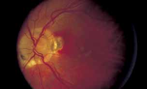
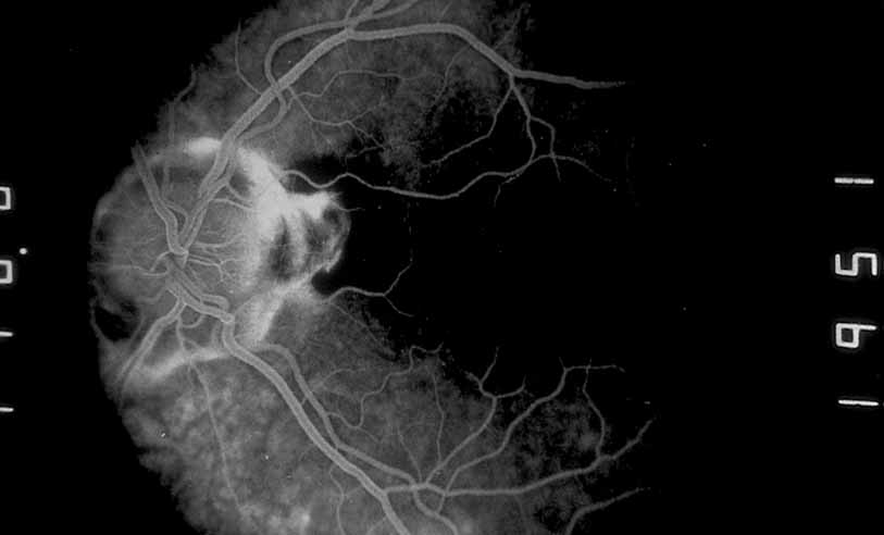
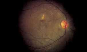
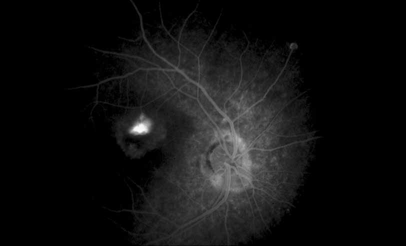
A variety of inflammations and infections, most notably presumed ocular histoplasmosis syndrome (see Figs. 37 and 38), serpiginous choroiditis, and multifocal choroiditis (see Figs. 39, 40, 41, and 42),1,89,90 are associated with the late complication of choroidal neovascularization. Probably the single most important application of fluorescein angiography in infectious and inflammatory diseases of the posterior segment is for the evaluation and potential treatment of this complication. In some instances, fluorescein angiography may be the single best method to determine whether visual loss seen late in the course of such disease as paramacular toxoplasmosis or serpiginous choroiditis is the result of secondary choroidal neovascularization or inflammatory disease recrudescence. In other diseases, such as presumed ocular histoplasmosis syndrome or multifocal choroiditis, choroidal neovascularization may occur early and be suspected on the basis of associated hemorrhage or lipid exudation. In some instances, even with the benefit of high-quality fluorescein angiography it may be difficult to distinguish between choroidal vasculitis and choroidal neovascularization superimposed on underlying choroiditis. We have recently identified a subgroup of young female patients, frequently darkly pigmented, who develop solitary zones of elevated choroiditis that have features of choroidal neovascularization. These appear to have a more indolent course than either idiopathic choroidal neovascularization or that associated with atypical multifocal choroiditis.

Fig. 37 Presumed ocular histoplasmosis. Color photograph showing subretinal hemorrhage adjacent to the optic disk.

Fig. 38 Presumed ocular histoplasmosis. Fluorescein angiography shows a small peri-papillary choroidal neovascular membrane.

Fig. 39 Multifocal choroiditis. Color photograph of the macula shows multiple lesions and an area suspicious for choroidal neovascularization.

Fig. 40 Multifocal choroiditis. Fluorescein angiography confirms the presence of a subfoveal choroidal neovascular membrane.

Fig. 41 Multifocal choroiditis. Color photograph shows multiple small choroidal lesions scattered throughout the posterior pole.

Fig. 42 Multifocal choroiditis. ICG angiography shows multiple hypofluorescent spots in the distribution of the choroidal lesions seen on color photography.

Back to Top
CONCLUSIONS
Fluorescein and IGC angiography are useful diagnostic tools in the evaluation of certain infections and inflammations of the posterior segment. Because of its biophysical properties, fluorescein acts as an image enhancer and also provides valuable information about the dynamic state of the blood-ocular barriers. Rarely, fluorescein or ICG angiography may result in a pathognomonic picture that convincingly establishes a diagnosis. More frequently, however, angiography provides useful information regarding the delineation of disease severity, mechanism of visual loss, and response to treatment. It enhances the ability of the ophthalmologist to distinguish between optic nerve, retinal, retinal vascular, pigment epithelial, and choroidal disease. It is especially useful in corroborating the presence of retinal or choroidal neovascularization or cystoid macular edema amenable to treatment, particularly in eyes with partially opacified media as a result of the intraocular inflammation.
Back to Top
REFERENCES

1. Gass JDM: Stereoscopic Atlas of Macular Diseases: Diagnosis and Treatment, 3rd ed. St. Louis: CV Mosby, 1987

2. Orth D: Color and Fluorescein Angiographic Atlas of Retinal Vascular Disorders. Baltimore: Williams & Wilkins, 1984

3. Ryan SJ: Retina, Vol 2, Medical Retina. St. Louis: CV Mosby, 1989

4. Yannuzzi LA, Gitter KA, Schatz H: The Macula: A Comprehensive Text and Atlas. Baltimore: Williams & Wilkins, 1979

5. Obana A, Kusumi M, Miki T: Indocyanine Green Angiographic Aspects of Multiple Evanescent White Dot Syndrome. Retina 16:97–104, 1996

6. Ie D, Glaser BM, Murphy RP, et al: Indocyanine Green Angiography in Multiple Evanescent White-Dot Syndrome. Am J Ophthalmol 117:7–12, 1994

7. Ikeda N, Ikeda T, Tano R, et al: Location of Lesions in Multiple Evanescent White Dot Syndrome and the Cause of the Hypofluorescent Spots Observed by Indocyanine Green Angiography. Graefe's Arch Clin Exp Ophthalmol 239:242–247, 2001

8. Dhaliwal RS, Maguire AM, Flower RW, et al: Acute Posterior Multifocal Placoid Pigment Epitheliopathy. Retina 13:317–325, 1993

9. Vadala M, Lodato G, Cillino S: Multifocal Choroiditis: Indocyanine Green Angiographic Features. Ophthalmologica 215:16–21, 2001

10. Okada AA, Mizusawa T, Sakai J, et al: Videofundoscopy and Videoangiography Using the Scanning Laser Ophthalmoscope in Vogt-Koyanagi-Harada Syndrome. Br J Ophthalmol 82:1175–1181, 1998

11. Oshima Y, Harino S, Hara Y, et al: Indocyanine Green Angiographic Findings in Vogt-Koyanagi-Harada Disease. Am J Ophthalmol 122:58–66, 1996

12. Yuzawa M, Kawamura A, Matsui M: Indocyanine Green Video-Angiographic Findings in Harada's Disease. Jpn J Ophthalmol 37:456–466, 1993

13. Matsuo T, Itami M, Shiraga F: Choroidopathy in Patients with Sarcoidosis Observed by Simultaneous Indocyanine Green and Fluorescein Angiography. Retina 20:16–21, 2000

14. Bozzoni-Pantaleoni F, Gharbiya M, Pirraglia M, et al: Indocyanine Green Angiographic Findings in Behcet Disease. Retina 21:230–236, 2001

15. Matsuo T, Sato Y, Shiraga F, et al: Choroidal Abnormalities in Behcet Disease Observed by Simultaneous Indocyanine Green and Fluorescein Angiography with Scanning Laser Ophthalmoscopy.

16. Arruga J, Valentines J, Mauri F, et al: Neuroretinitis in acquired syphilis. Ophthalmology 92:262, 1985

17. Cunha De Souza E, Jalkh AE, Trempe CL, et al: Unusual central chorioretinitis as the first manifestation of early secondary syphilis. Am J Ophthalmol 105:271, 1988

18. DeLuise VP, Clark SW, Smith JL: Syphilitic retinal detachment and uveal effusion. Am J Ophthalmol 94:757, 1982

19. Halperin LS, Lewis H, Blumenkranz MS, et al: Choroidal neovascular membrane and other chorioretinal complications of acquired syphilis. Am J Ophthalmol 108:554, 1989

20. Mendelsohn AD, Jampol LM: Syphilitic retinitis: A cause of necrotizing retinitis. Retina 4:221, 1984

21. Morgan CM, Webb RM, O'Connor GR: Atypical syphilitic chorioretinitis and vasculitis. Retina 4:225, 1984

22. Blumenkranz MS, Gass JDM, Clarkson JG: Atypical serpiginous choroiditis. Arch Ophthalmol 100:1773, 1982

23. Broekhuyse RM, van Herck M, Pinckers AJ, et al: Immune responsiveness to retinal S-antigen and opsin in serpiginous choroiditis and other retinal disease. Doc Ophthalmol 69:83, 1988

24. Exp Ophthalmol 219:131, 1982

25. Hardy RA, Schatz H: Macular geographic helicoid choroidopathy. Arch Ophthalmol 105:1237, 1987

26. Mansour AM, Jampol LM, Packo L, et al: Macular serpiginous choroiditis. Retina 8:125, 1988

27. Schatz H, Maumenee AE, Patz A: Geographic helicoid peripapillary choroidopathy: Clinical presentations and fluorescein angiographic findings. Trans Am Acad Ophthalmol Otolaryngol 78:747, 1974

28. Wojno T, Meredith TA: Unusual findings in serpiginous choroiditis. Am J Ophthalmol 94:650, 1982

29. Wu JS, Lewis H, Fine SL, et al: Clinicopathologic findings in a patient with serpiginous choroiditis and treated choroi-dal neovascularization. Retina 9:292, 1989

30. Friberg TR: Serpiginous choroiditis with branch vein occlusion and bilateral periphlebitis: Case report. Arch Ophthalmol 106:585, 1988

31. Eichenbaum JW, Friedman AH, Mamelok AE: A clinical and histopathological review of intermediate uveitis (pars planitis). Bull NY Acad Med 64:164, 1988

32. Henderly DE, Haymond RS, Rao NA, Smith RE: The significance of the pars plana exudate in pars planitis. Am J Ophthalmol 103:669, 1987

33. Wetzig RP, Chan CC, Nussenblatt RB, et al: Clinical and immunopathologic studies of pars planitis in a family. Br J Ophthalmol 72:5, 1988

34. Brod RD: Presumed sarcoid choroidopathy mimicking birdshot retinochoroidopathy. Am J Ophthalmol 109:357, 1990

35. Campo RV, Aaberg TM: Choroidal granuloma in sarcoidosis. Am J Ophthalmol 97:419, 1984

36. Duker JS, Brown GC, McNamara JA: Proliferative sarcoid retinopathy. Ophthalmology 95:1680, 1988

37. Franceschetti A, Babel J: La choriorétinité en taches de bougie: Manifestations de la maladie de Besnier-Boech. Ophthalmologica 118:701, 1949

38. Marcus DF, Bovino JA, Burton TC: Sarcoid granuloma of the choroid. Ophthalmology 89:1326, 1982

39. Mizuno K, Takahashi J: Sarcoid cyclitis. Ophthalmology 93:511, 1986

40. Spalton DJ, Sanders MD: Fundus changes in histologically confirmed sarcoidosis. Br J Ophthalmol 65:348, 1981

41. Chan CC, Palestine AG, Kuwabara T, Nussenblatt RB: Immunopathologic study of Vogt-Koyanagi-Harada syndrome. Am J Ophthalmol 105:607, 1988

42. Albert DM, Diaz-Rohena R: A historical review of sympathetic ophthalmia and its epidemiology. Surv Ophthalmol 34:1, 1989

43. Carney MD, Tessler HH, Peyman GA, et al: Sympathetic ophthalmia and subretinal neovascularization. Ann Ophthalmol 22:184, 1990

44. Jennings T, Tessler HH: Twenty cases of sympathetic ophthalmia. Br J Ophthalmol 73:140, 1989

45. Maisel JM, Vorwerk PA: Sympathetic uveitis after giant tear repair. Retina 9:122, 1989

46. Tessler HH, Jennings T: High-dose short-term chlorambucil for intractable sympathetic ophthalmia and Behçet's disease. Br J Ophthalmol 74:353, 1990

47. Blumenkranz MS, Kaplan HJ, Clarkson JG, et al: Acute multifocal hemorrhagic retinal vasculitis. Ophthalmology 95:1663, 1988

48. Bertram B, Wolf S, Hof A, et al: Rheologic findings in patients with Eale's disease. Klin Monatsbl Augenheilkd 195:254, 1989

49. Gordon MF, Coyle PK, Golub B: Eale's disease presenting as stroke in the young adult. Ann Neurol 24:264, 1988

50. Magargal LE, Walsh AW, Magargal HO, et al: Treatment of Eale's disease with scatter laser photocoagulation. Ann Ophthalmol 21:300, 1989

51. Moyenin P, Bonnet M, Grange JD: Eale's syndrome and photocoagulation: A propos of 29 cases. Bull Soc Ophthalmol Fr 87:383, 1987

52. Riss I, Le-Rebeller MJ, Tapiero B: Extensive fibrovascular proliferations in Eale's disease. Bull Soc Ophthalmol Fr 86:1549, 1555, 1986

53. Blumenkranz M, Clarkson J, Culbertson WW, et al: Visual results and complications after retinal reattachment in the acute retinal necrosis syndrome: The influence of operative technique. Retina 9:170, 1989

54. Blumenkranz MS, Clarkson J, Culbertson WW, et al: Vitrectomy for retinal detachment associated with acute retinal necrosis. Am J Ophthalmol 106:426, 1988

55. Browning DJ, Blumenkranz MS, Culbertson WW, et al: Association of varicella zoster dermatitis with acute retinal necrosis syndrome. Ophthalmology 94:602, 1987

56. Clarkson JG, Blumenkranz MS, Culbertson WW, et al: Retinal detachment following the acute retinal necrosis syndrome. Ophthalmology 91:1665, 1984

57. Culbertson WW, Blumenkranz MS, Haines H, et al: The acute retinal necrosis syndrome: II. Histopathology and etiology. Ophthalmology 89:1317, 1982

58. Culbertson WW, Blumenkranz MS, Pepose JS, et al: Varicella zoster virus is a cause of the acute retinal necrosis syndrome. Ophthalmology 93:559, 1986

59. Bisher JP, Lewis ML, Blumenkranz MS: The acute retinal necrosis syndrome: I. Clinical manifestations. Ophthalmology 89:1309, 1982

60. Han DP, Lewis H, Williams GA, et al: Laser photocoagulation in the acute retinal necrosis syndrome. Arch Ophthalmol 105:1051, 1987

61. Holland GN, Cornell PJ, Park MS, et al: An association between acute retinal necrosis syndrome and HLA-DQw7 and phenotype Bw62, DR4. Am J Ophthalmol 108:370, 1989

62. Immunen I, Laatikainen L, Linnanvuori K: Acute retinal necrosis syndrome treated with vitrectomy and intravenous acyclovir. Acta Ophthalmol 67:106, 1989

63. Lewis ML, Culbertson WW, Post JD, et al: Herpes simplex virus type I:. A cause of the acute retinal necrosis syndrome. Ophthalmology 96:875, 1989

64. Ludwig IH, Zegarra H, Zakov ZN: The acute retinal necrosis syndrome: Possible herpes simplex retinitis. Ophthalmology 91:1659, 1984

65. Matsuo T, Koyama M, Matsuo N: Acute retinal necrosis as a novel complication of chickenpox in adults. Br J Ophthalmol 74:443, 1990

66. Matsuo T, Ohno A, Matsuo N: Acute retinal necrosis syndrome following chickenpox in pregnant woman. Jpn J Ophthalmol 32:70, 1988

67. Saga U, Ozawa H, Soshi S, et al: Acute retinal necrosis (Kirisawa's uveitis). Jpn J Ophthalmol 27:353, 1983

68. Sergott RC, Belmont JB, Savino PJ, et al: Optic nerve involvement in the acute retinal necrosis syndrome. Arch Ophthalmol 103:1160, 1985

69. Sternberg P Jr , Know DL, Finkelstein D, et al: Acute retinal necrosis syndrome. Retina 2:145, 1982

70. Wang CL, Kaplan HJ, Waldrep JC, et al: Retinal neovascularization associated with acute retinal necrosis. Retina 3:249, 1983

71. Yeo JH, Pepose JS, Stewart JA: Acute retinal necrosis syndrome following herpes zoster dermatitis. Ophthalmology 93:1418, 1986

72. Schilling H, Bornfeld N, Windeck R, et al: Cystostatic and immunosuppressive treatment of ocular Behçet's syndrome. Klin Monatsbl Augenheilkd 196:62, 1990

73. Sugihara I: Measurement of retinal circulation in uveitis of Behçet's and Harada's disease. Nippon Geka Gakkai Zasshi 93:705, 1989

74. Wakefield D, McCluskey P: Behçet's syndrome: Ocular features in Australian population. Aust NZ J Ophthalmol 18:129, 1990

75. Parke DW, Font RL: Diffuse toxoplasmic retinochoroiditis in a patient with AIDS. Arch Ophthalmol 104:571, 1986

76. Brown GC, Brown RH, Brown MM: Peripheral proliferative retinopathies. Int Ophthalmol 11:41, 1987

77. James CR, Jacobs PM, Leaver PK, McLeod D: Closed microsurgery for the sequelae of neovascularization from veno-occlusive retinopathies. Eye l(pt 1) :106, 1987

78. Regan CD, Foster CS: Retinal vascular diseases: Clinical presentation and diagnosis. Int Ophthalmol Clin. 26:25, 1986

79. Jampol LM, Sieving PA, Pugh D, et al: Multiple evanescent white dot syndrome. Arch Ophthalmol 102:671, 1984

80. Charteris DG, Khanna V, Dhillon B: Acute posterior multifocal placoid pigment epitheliopathy complicated by central retinal vein occlusion. Br J Ophthalmol 73:765, 1989

81. Foulds WS, Damato BE: Investigation and prognosis in the retinal pigment epitheliopathies. Aust NZ J Ophthalmol 14:301, 1986

82. Gass JDM: Acute posterior multifocal placoid pigment epitheliopathy. Arch Ophthalmol 80:177, 1968

83. Isashiki M, Koide H, Yamashita T, Ohba N: Acute posterior multifocal placoid pigment epitheliopathy associated with diffuse retinal vasculitis and late hemorrhagic macular detachment. Br J Ophthalmol 70:255, 1986

84. Kersten DH, Lessell S, Carlow TJ: Acute posterior multifocal placoid pigment epitheliopathy and late-onset meningo-encephalitis. Ophthalmology 94:393, 1987

85. Williams DF, Mieler WF: Long-term follow-up of acute multifocal posterior placoid pigment epitheliopathy. Br J Ophthalmol 73:985, 1989

86. Schlaegel TF: Presumed histoplastic choroiditis. Am J Ophthalmol 63:919, 1967

87. Pepose JS, Holland GN, Nestor MS, et al: Acquired immune deficiency syndrome: Pathogenic mechanisms of ocular disease. Ophthalmology 92:472, 1985

88. Schuman JS, Friedmari AH: Retinal manifestations of the acquired immune deficiency syndrome (AIDS): Cytomegalovirus, Candida albicans, Cryptococcus, toxoplasmosis, and Pneumocystis carinii. Trans Ophthalmol Soc UK 103(pt 2):177, 1983

89. Cantrill HL, Folk JC: Multifocal choroiditis associated with progressive subretinal fibrosis. Am J Ophthalmol 101:170, 1986

90. Dreyer RF, Gass JDM: Multifocal choroiditis and panuveitis: A syndrome that mimics ocular histoplasmosis. Arch Ophthalmol 102:1776, 1984

Back to Top