|
|
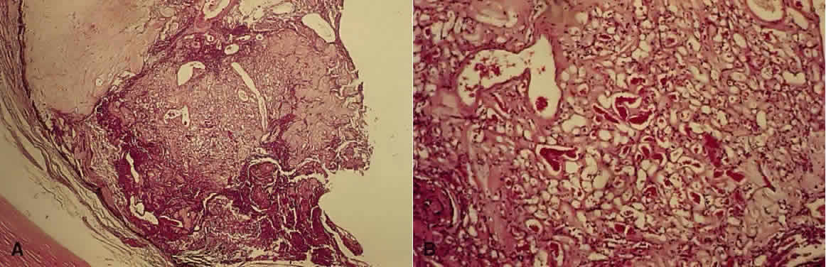
| Fig. 4. Retinal hemangioblastoma. A. Low-power view shows a hemangioblastoma at the ora serrata with disruption of normal architecture. The ciliary epithelium has proliferated beneath the hemangioblastoma (H&E, × 7.8). B. Higher-power view shows the vascular channels (H&E, × 31). |