|
|
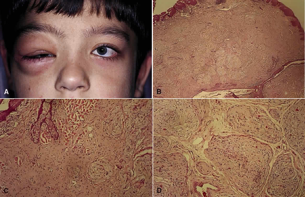
| Fig. 7. Neurofibromatosis type 1. A. Clinical view of neurofibroma involving the right lids (H&E, × 7.8). B. Low-power view showing diffuse involvement of the dermis (H&E, × 31). C. Higher-power view shows infiltration and separation of striated muscle fibers (H&E, × 31). D. In another area are bundles of proliferated neural elements. |