|
|
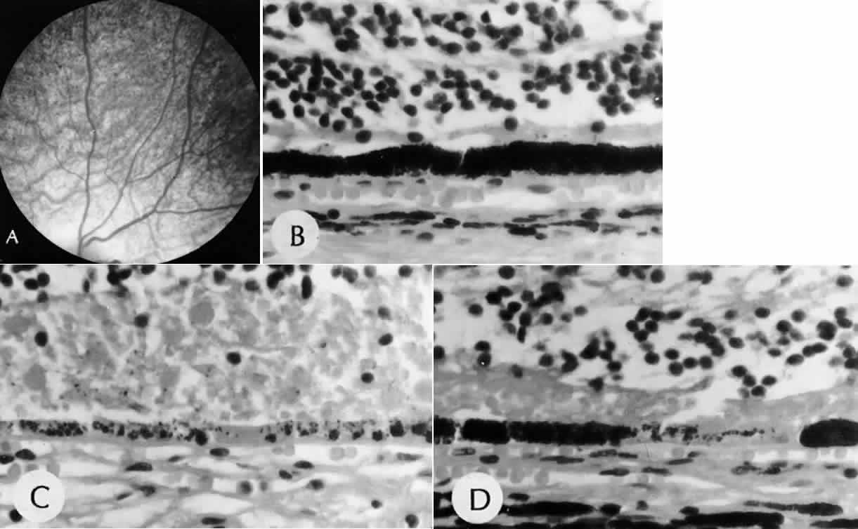
| Fig. 17. Rubella. A. Fundus picture shows mottled “salt-and-pepper” appearance (SEI 79-37). B through D. All from same eye. Retinal pigment epithelium shows areas of hyperpigmentation (B), hypopigmentation (C), and alternating areas of hypo- and hyperpigmentation (D) (B, C, and D, H&E, × 630 [SEI 79-38, 79-39, and 79-40)]. (Modified from Yanoff M: The retina in rubella. In Tasman W [ed]: Retinal Diseases in Children, p 223. New York, Harper & Row, 1971. |