|
|
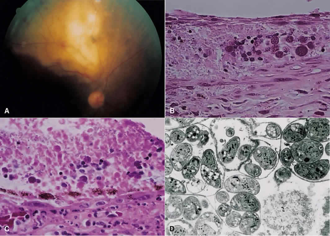
| Fig. 13 A. Right fundus from patient with the acquired immunodeficiency syndrome showing a large area of necrotizing retinitis (pigmentation within area of necrosis, possibly representing previous site of activity) with hemorrhage, which, on histopathologic analysis (B and C), revealed retinal necrosis and the presence of cysts consistent with Toxoplasma gondii (hematoxylin and eosin; B × 200, C × 500). D. Electron microscopy shows tachyzoites within cysts. |