|
|
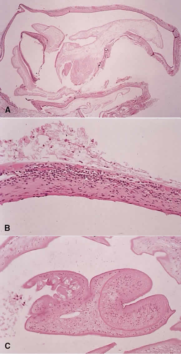
| Fig. 14. Echinococcus. A. Low-power photo of a hydatid cyst removed from an orbit of an 8-year-old child. B. Higher power of the wall of the cyst reveals inflammation with abundant eosinophils. C. The scolex and hooklets of the larval form within the cyst lumen. |