
|
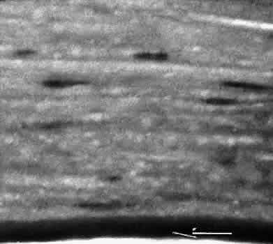
| Fig. 36. Light micrograph of a corneal specimen removed because of bullous keratopathy. Descemet's membrane (arrow) is of normal caliber and character for the patient's age. No endothelial cells can be identified at the light microscopic level. Because of the normal caliber of Descemet's membrane, the endothelial cell change appears to be of short duration and is attributed to problems associated with pseudophakia (pseudophakic bullous keratopathy). (Periodic acid-Schiff stain; × 100.) |