
|
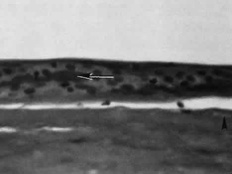
| Fig. 37. Light micrograph of corneal epithelium in a case of surgically treated pseudophakic bullous keratopathy. Repeated erosion and healing of the epithelial layer has resulted in the formation of an intraepithelial basement membrane (arrow). There is also an area ofsubepithelial bullous formation and a small area of subepithelial fibrous tissue formation (arrowhead). The epithelial changes in Fuchs' endothelial dystrophy are identical to pseudophakic bullous keratopathy. (Periodic acid-Schiff stain; × 16.) |