|
|
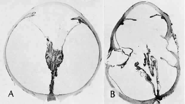
| Fig. 42. Two cases of retinal detachment following cataract extraction. A. Retinal detachment was identified 4 weeks after cataract extraction. Fixed retinal folds indicate that the situation is inoperable, so no surgical repair was attempted. The anterior chamber angle has become occluded because of neovascularization associated with ischemic retina. B. Retinal detachment was identified 5 weeks after cataract extraction. Two attempts at surgical repair failed. The large equatorial cystic spaces indicated the presence of a scleral-buckling element. The actual material of the sponge and buckle has been lost during tissue processing. The cystic nature of the detached retinal tissue indicates that there was an extended time between the last retinal reattachment attempt and enucleation. In this case, peripheral anterior synechiae are present. The indication for enucleation was most likely a blind painful eye due to secondary glaucoma and reactive uveitis. (Hematoxylin-eosin stain; × 3.) |