
|
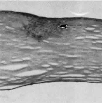
| Fig. 52. Light micrograph of a corneal specimen removed for complications of radial keratotomy 10 months postoperatively. A facet of epithelial cells remains in the anterior portion of the wound (arrow), indicating that the wound-healing process in this case is incomplete. The character of the wound also suggests that there is incomplete return of tensile strength in this area. (Hematoxylin-eosin stain; × 25.) |