|
|
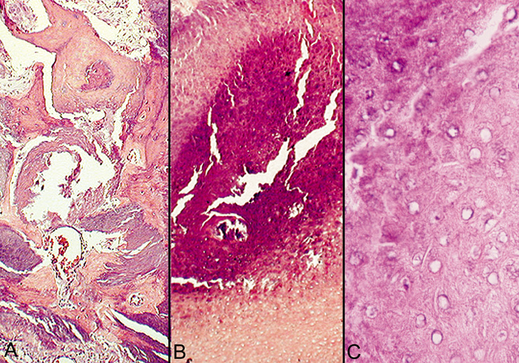
| Fig. 23. Pilomatrixoma—A. Low-power photomicrograph demonstrating immature bone formation and proliferation of epithelial cells (hematoxylin and eosin stain). B. Higher-power view showing an area of calcification within the tumor (ematoxylin and eosin stain). C. High-power photomicrograph showing the transition from basophilic epithelial cells (upper left) to “shadow cells“ in the lower right (hematoxylin and eosin stain). (Photos courtesy of William Morris, M.D.) |