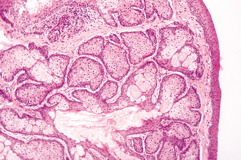
Fig. 31.
Sebaceous Hyperplasia—Low-power photomicrograph showing an enlarged sebaceous gland with many lobules and a dilated central duct (hematoxylin and eosin stain). (Photo courtesy of William Morris, M.D.)