|
|
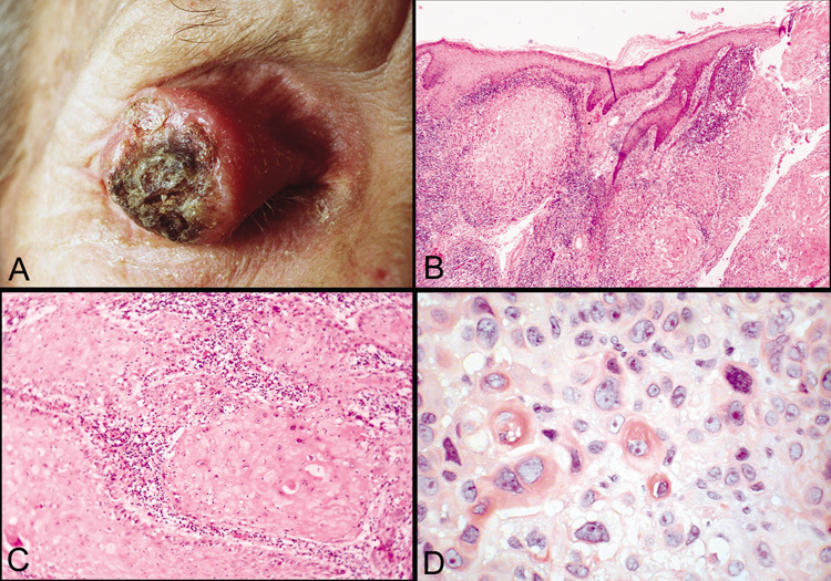
| Fig. 46. Squamous cell carcinoma—A. Clinical photograph of large squamous cell carcinoma on the upper eyelid. B. Low-power photomicrograph of tumor demonstrating eosinophilic squamous cells that have invaded the dermis and subcutaneous tissue. (hematoxylin and eosin stain). C. Higher-power photomicrograph showing lobules of invading tumor cells (hematoxylin and eosin stain). D. High-power photomicrograph illustrating dyskeratotic and atypical cells within tumor (hematoxylin and eosin stain). (Photos courtesy of William Morris, M.D.) |