
|
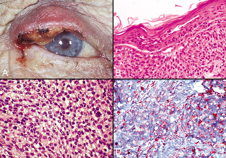
| Fig. 48. Sebaceous Carcinoma—A. Clinical photograph of patient with sebaceous carcinoma. B. Photomicrograph showing pagetoid spread of tumor cells within the epithelium (hematoxylin and eosin stain). C. High-power photomicrograph demonstrating foamy cytoplasm typical of this tumor (hematoxylin and eosin stain). D. Oil Red O stain identifies the lipid material produced by the tumor cells (Oil Red O stain). (Photos courtesy of William Morris, M.D.) |