|
|
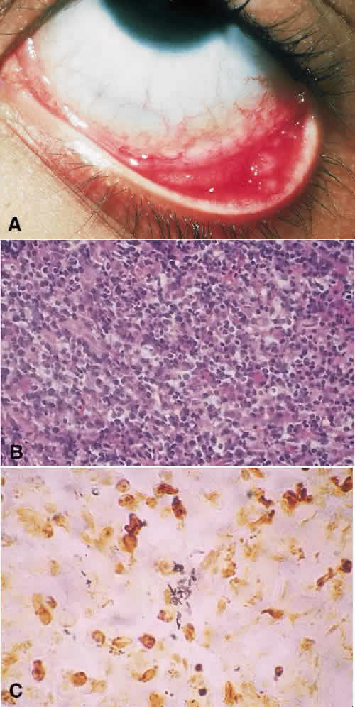
| Fig. 7. A. Clinical photograph of papillary conjunctivitis. Ipsilateral enlargement of the preauricular lymph nodes is consistent with Parinaud oculoglandular syndrome. B. Histopathology shows a dense infiltrate of lymphocytes and epithelioid histiocytes. C. Warthin—Starry stain shows numerous pleomorphic bacilli consistent with Rochalimaea henselae. |