|
|
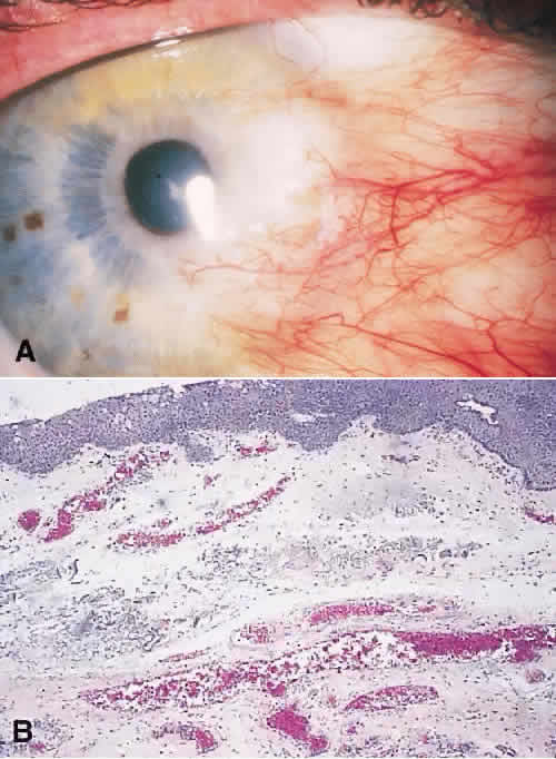
| Fig. 11. A. Clinical photograph of pterygium shows localized, yellowish gray, elevated, vascularized lesion of the conjunctiva extending onto the peripheral cornea. B. Histopathology shows thickened epithelium. Amorphous eosinophilic staining, hyalinized material, and numerous vessels are identified in the substantia propria of the conjunctiva. |