|
|
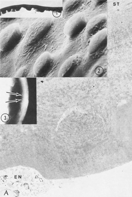
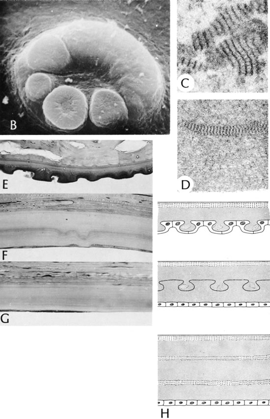
| Fig. 43. Cornea guttata. A. A full-thickness wart composed of a mixture of banded basement membrane (both 500 and 1000 angstrom), homogeneous basement membrane, and filamentous basement membrane. The debris of some remaining endothelium is present (EN). Wart-like thickening on Descemet's membrane in inset 1 (cornea guttata clinically—arrows in inset 3) is seen better by scanning electron microscopy in inset 2. Many warts are anvil shaped. ST, corneal stroma. B. Scanning electron micrograph of confluent warts. The warts often present in a variety of shapes. Banded basement membrane of 1000 angstroms (C) and 500 angstroms (D) variety in the wart. E. A typical wartlike configuration of Descemet's membrane. F. Warts are buried deep within the thickened Descemet's membrane. G. Uniformly thickened Descemet's membrane without evidence of wart formation. H. The drawing illustrates the three basic patterns of thickening of Descemet's membrane in cornea guttata. The dotted, white areas represent regions of banded basement membrane. (A, main figure, ×7200; inset 1, periodic acid–Schiff, ×350 [Armed Forces Institute of Pathology (AFIP) Neg. 77-3456]; inset 2, ×2000; inset 3, clinical, B, ×4500; C, ×27,000; D, ×30,000; E, F, and G, PD, ×300 [AFIP Negs. 77-3556, 77-3557, and 77-4176]; H, drawing) |