|
|
|
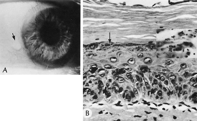
Fig. 45.
Bitot's spot. A. The clinical appearance (arrow). B. The conjunctival epithelium shows hyperkeratosis. Note the prominent granular layer (arrow). Pleomorphic gram-positive rods, compatible with diphtheroids, are clustered around the keratin debris (inset). (Courtesy of SEI Photoarchives.) ( |