
|
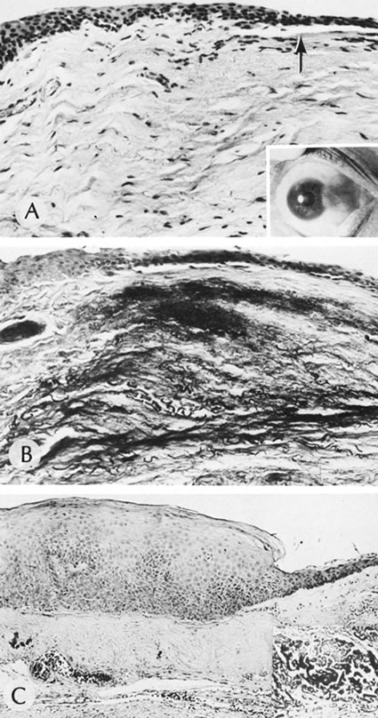
| Fig. 47. Pterygium. A. Basophilic degeneration in the subepithelial tissue. Dissolution of Bowman's membrane can be seen (arrow). The clinical appearance of pterygium is shown in the inset. B. An area of basophilic degeneration stains deeply with a stain for elastic tissue. C. Overlying epithelium shows mild dysplastic changes.The subepithelial tissue stains positively for elastic tissue, as shown in the inset. (Courtesy of SEI Photoarchives.) |