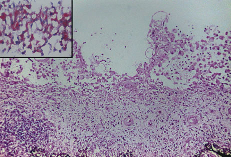
Fig. 3.
Juvenile xanthogranuloma. The iris is infiltrated by histiocytes, which form nodular aggregates on the anterior surface of the iris. (Hemotoxylin-eosin ×25.) Inset (×200) shows oil red O-positive histiocytes.