|
|
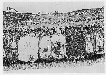
| Fig. 9. Hemorrhage within the nerve fiber layer (arrow) tends to “track” along the nerve fibers, producing a flame-shaped hemorrhage observed clinically. An accumulation of blood in the potential space between the internal limiting membrane (ilm) and the nerve fiber layer produces submembranous intraretinal hemorrhage, which is generally restrained from entering the vitreous compartment by the strength of the overlying thick basement membrane. Pockets of blood accumulating within the bipolar cell layer or between Henle's fibers (photoreceptor axons) of the extramacular outer plexiform layer produce the dot and blot hemorrhages seen clinically. (H&E, × 115) |