|
|
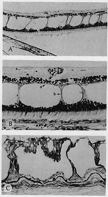
| Fig. 13. Typical microcystoid degeneration. A. Spaces or channels lie initially within outer plexiform layer (i.e., between compressed bundles of photoreceptor axons and the remains of Müller glial cells). B. Higher magnification shows that the middle limiting membrane (a) limits the inner boundaries, the glial-neuronal columns (b) limit the lateral boundaries, and the photoreceptor cell bodies (outer nuclear layer) and the external limiting membrane (c) limit the outer boundaries of the microcystoid channels. C. Diphosphopyridine nucleotide (DPNH) diaphorase (nitro-blue tetrazolium [BT] method). The glial-neuronal columns show a dense precipitate of formazan, signifying the presence of the cytochrome oxidase system. (A, H&E, ×135; B, H&E, ×300; C, nitro-BT, × 125) |